XB-IMG-75178
Xenbase Image ID: 75178
|
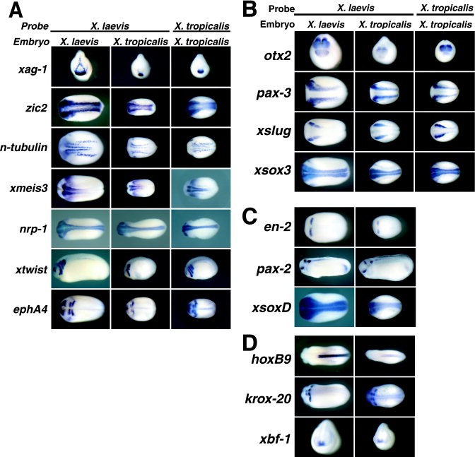
Figure 2. In situ hybridizations were done for ectodermally expressed genes in Xenopus laevis and X. tropicalis embryos. For all panels, X. laevis probes used on X. laevis embryos (first column) and X. tropicalis embryos (middle column) are shown. A,B:X. tropicalis probes used on X. tropicalis embryos (right column). Some X. laevis probes were found to work well on X. tropicalis embryos (A,C), whereas others worked weakly (B,D). For xag-1, otx2, and xbf-1, anterior views (dorsal side up) are shown for stage 22, late neurula, and stage 24 embryos, respectively. For zic2, xmeis3, ephA4, pax-3, xslug, xsox3, engrailed-2 (en-2), and xsoxD, dorsal views of late neurula staged embryos (anterior to the left) are shown. For neural-specific β-tubulin (n-tubulin), dorsal views of early neurula staged embryos (anterior to the left) are shown. For nrp-1, hoxB9, and krox-20, dorsal views of stage 24â25 embryos (anterior to the left) are shown. For xtwist and pax-2, lateral views are shown (anterior to the left) of stage 24 and stage 27 embryos, respectively.Download figure to PowerPoint Image published in: Khokha MK et al. (2002) Copyright © 2002. Image reproduced with permission of the Publisher, John Wiley & Sons. Larger Image Printer Friendly View |