XB-IMG-75210
Xenbase Image ID: 75210
|
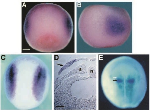
Fig. 2. Whole-mount in situ hybridization analysis of Pax-3 expression in Xenopus embryos. (A) Dorsovegetal and, (B) lateral view at stage 11.5 showing that Pax-3 is expressed in distinct lateral domains of the presumptive neural plate. (C) Dorsal view at stage 12 showing the refinement of Pax-3 expression to lateral domains of the neural plate during convergence and extension. (D) Transverse paraffin section of a stage 16 embryo, showing that Pax-3 expression is restricted to the lateral neural plate (arrow), overlying somitic and lateral plate mesoderm. (E) Dorsoanterior view of a stage 18 embryo hybridized with Pax-3 (light blue, rostral extent indicated by a white arrow) and en-2 (purple, indicated by a black arrow) showing that Pax-3 expression extends just rostral to the mb-hb boundary. n, notochord; s, somite. Scale bars, (A-C, E) 200 μm; (D) 100 μm. Image published in: Bang AG et al. (1997) Copyright © 1997. Image reproduced with permission of the Publisher and the copyright holder. This is an Open Access article distributed under the terms of the Creative Commons Attribution License.
Image source: Published Larger Image Printer Friendly View |