Click here to close Hello! We notice that you are using Internet Explorer, which is not supported by Xenbase and may cause the site to display incorrectly. We suggest using a current version of Chrome, FireFox, or Safari.
XB-IMG-75447

Xenbase Image ID: 75447

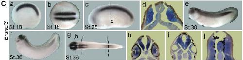
Fig. 3. Embryonic expression of the X. laevis Brunol genes at the indicated stages.(C) Brunol3, (a,c,e,f), lateral views, anterior to the left; (b,g), dorsal views, anterior to the left.

Image published in: Wu J et al. (2010)

Copyright © 2010. Reproduced with permission of the Publisher, University of the Basque Country Press.

GeneSynonymsSpeciesStage(s)Tissue
celf2.Lbrunol3, celf2-a, celf2-b, cugbp2, etr-3, etr3, naporX. laevisThroughout NF stage 18presomitic mesoderm
celf2.Lbrunol3, celf2-a, celf2-b, cugbp2, etr-3, etr3, naporX. laevisThroughout NF stage 25presomitic mesoderm
celf2.Lbrunol3, celf2-a, celf2-b, cugbp2, etr-3, etr3, naporX. laevisThroughout NF stage 29 and 30hindbrain
midbrain
somite
presomitic mesoderm
optic vesicle
spinal cord
cement gland
fin
celf2.Lbrunol3, celf2-a, celf2-b, cugbp2, etr-3, etr3, naporX. laevisThroughout NF stage 35 and 36somite
presomitic mesoderm
hindbrain
midbrain
otic vesicle
posterior neural tube
pharyngeal arch
facial epibranchial placode
lens
retina

Image source: Published

Larger Image
Printer Friendly View

Return to previous page