XB-IMG-76185
Xenbase Image ID: 76185
|
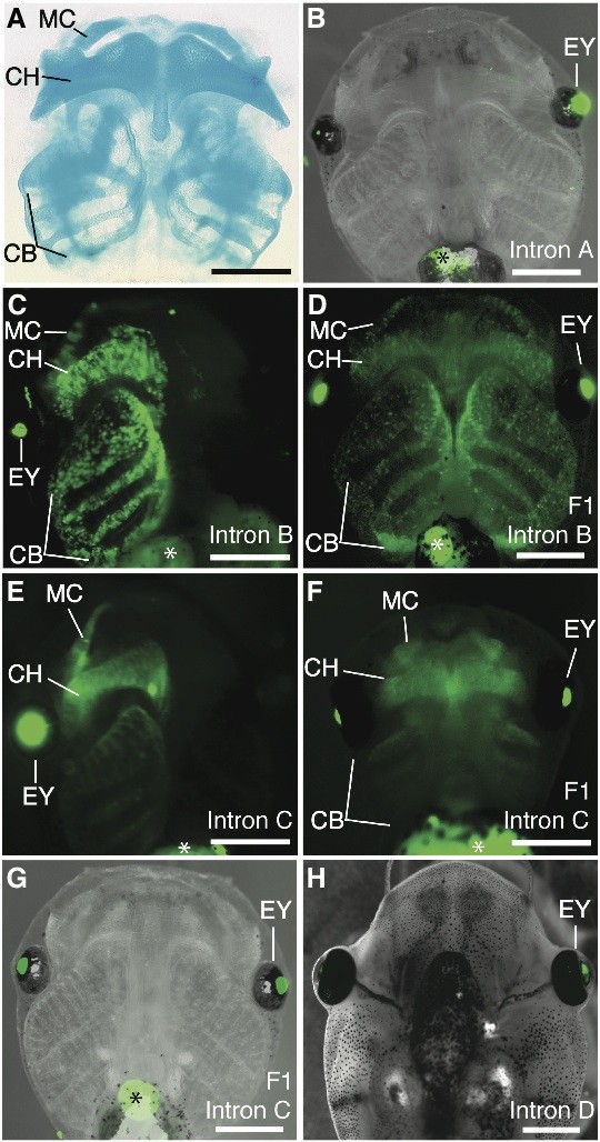
Fig. 3. Distribution of EGFP in transgenic Xenopus laevis tadpoles. (A) Ventral views, anterior to the top. (H) Dorsal view, anterior to the top. (A) Cleared-and-stained tadpole showing Meckel (MC), ceratohyal (CH) and ceratobranchial (CB) cartilages stained with Alcian blue (stage 42). All other images show EGFP/GFP expression in living tadpoles. (B) Transient transgenic tadpole with a reporter carrying the intronic region A (stage 48). EGFP is not seen in the cartilaginous skeleton of any tadpole carrying reporter construct A. GFP expression is apparent in the lens of the left eye (EY), under the control of the gamma-crystallin promoter. (C) Transient alf-transgenictadpole with a reporter carrying the intronic region B (stage 42). Unilateral transgene expression is apparent in the same cartilages labeled in (A), along with GFP expression in the eye. (D) An F1 transgenic tadpole (stage 48), bred from a half-transgenic mother carrying construct B. (E) Transient half-transgenic tadpole carrying a reporter with the C construct (stage 42) also exhibits unilateral cartilagi- nous expression, although expression is weaker than that seen in tadpoles carrying the B construct. (F) An F1 transgenic tadpole (stage 40), bred from a half-transgenic mother carrying construct C. Early expression of EGFP is strongest as the cartilages are forming. (G) Stage 48 of the same tadpole in (F). None of the tadpole cartilages continues to express EGFP under construct C during the later larval stages. However, cartilagi- nous expression of EGFP increases again during the metamorphic formation of adult cartilages (Fig. 4). (H) Merged bright-field and fluores- cent image of a transgenic tadpole carrying a reporter with the smaller intronic region D (stage 48). This region does not drive cartilaginous EGFP expression during any stage in any injected tadpole. Auto-fluorescence is visible in the yolk of stage 402 individuals (asterisks in C,E,F) and in the gallbladder of stage-48 tadpoles (asterisks in B,D,G). Scale bar, 1 mm. Image published in: Kerney R et al. (2010) Copyright © 2010. Reproduced with permission of the Publisher, University of the Basque Country Press. Larger Image Printer Friendly View |