XB-IMG-76983
Xenbase Image ID: 76983
|
||||||||||||||||||||
|
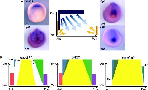
Fig. 10. Model of signalling input into the LPM of the neurula stage Xenopus embryo. A high level of RA signalling input is suggested by the expression domain of raldh2 in the anterior and dorsal LPM (A). Conversely, Fgf signalling is proposed in the anterior-ventral and posterior-ventral LPM, suggested by the expression domains of fgf8 in the anterior, and fgf4 and fgf8 in the posterior pole of the embryo. Dark blue and yellow bars represent the expression of raldh2 and fgf ligands respectively, while blue and yellow arrows depict the proposed area which RA (blue) and fgf (yellow) are required for normal patterning. The model depicts a left lateral view of the LPM. Ant: anterior, Dor: dorsal, Lat: lateral view, Pos: posterior, Ven: ventral. (B) Diagrammatical representation of LPM expression domain response to decrease in either RA (left) or Fgf (right) signalling when compared with the DMSO control (centre). The anterior-dorsal and middle LPM domains require RA signalling for their full expression domain. When embryos are treated with the RA antagonist the anterior-dorsal domain is restricted (a) and the middle LPM domain is contracted (b). When Fgf signalling is inhibited the anterior-dorsal domain is expanded ventrally (c) and the posterior domains are severely restricted (d), indicating that the anterior-ventral and posterior LPM domains are dependant on Fgf signalling. Red: nkx2.5, blue: foxf1, yellow: hand1, green: sall3, purple: Xbra. Image published in: Deimling SJ and Drysdale TA (2011) Copyright © 2011. Image reproduced with permission of the Publisher, Elsevier B. V.
Image source: Published Larger Image Printer Friendly View |