XB-IMG-77078
Xenbase Image ID: 77078
|
|
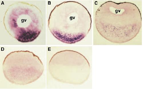
Fig. 3. Localisation of maternal Apod mRNA throughout oogenesis and early cleavage stages in Xenopus. Apod mRNA was visualised by in situ hybridisation to previously sectioned material using DIG-labelled antisense riboprobes (A-D) or sense probes as negative controls (E). Cells are orientated such that the animal pole is uppermost. (A) Stage 3 oocyte showing presence of Apod mRNA in both the vegetal cortex and yolk mass. (B) Stage 4 oocyte. (C) Mature stage 6 oocyte showing the particulate localisation of Apod mRNA in the vegetal yolk mass. The mRNA is now localised in a more subequatorial position. (D) Egg showing more diffuse localisation of Apod mRNA, still distributed in the vegetal yolk mass. (E) Egg hybridised with a sense probe showing very faint non-specific staining throughout the animal hemisphere only. A and B are shown at a greater magnification than C-E for convenient comparison. (gv, germinal vesicle) Image published in: Stennard F et al. (1996) Copyright © 1996. Image reproduced with permission of the publisher and the copyright holder. This is an Open Access article distributed under the terms of the Creative Commons Attribution License. Larger Image Printer Friendly View |