XB-IMG-77241
Xenbase Image ID: 77241
|
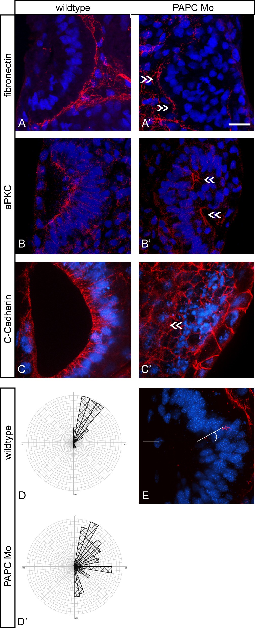
Figure 4. Immunostaining of otic epithelia. (A, A', B, B', C, C') ISH for Tbx2 was performed at stage 26. Embryos were then sliced and immunostained for different epithelial markers, like (A, A') fibronectin (n = 23), (B, B') aPKC (n = 19) and (C, C') C-Cadherin (n = 23) in red. Nuclei were stained with DAPI in blue. The otocyst region could be identified in the sections by Tbx2 expression. (A', B', C') Injected otocysts show epithelial disorganization indicated through inaccurate nuclei alignment and displaced epithelial markers (arrows). (D, D') Rose diagram to highlight the orientation of cells in wildtype (D) compared to morphant (D') otocyst region. Wildtype otocysts show cells orientated mostly in angles between 0 and 45hile morphant otocysts display angles between 0 and 180 (E) Demonstration of the angle measurements: orientation of DAPI stained nuclei in relation to a horizontal median through the otocyst. Scale bar 20 μm. Image published in: Jung B et al. (2011) Copyright ©2011 Jung et al; licensee BioMed Central Ltd. This image is reproduced with permission of the journal and the copyright holder. This is an open-access article distributed under the terms of the Creative Commons Attribution license
Image source: Published Larger Image Printer Friendly View |