XB-IMG-78293
Xenbase Image ID: 78293
|
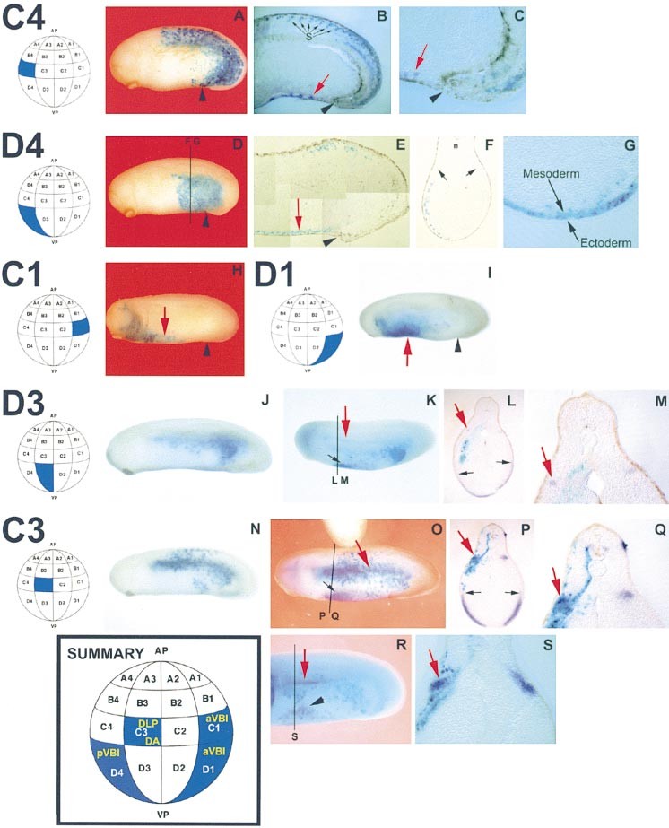
Figure 2. Blastomeres Contributing to Primitive and Definitive Blood Compartments in the Xenopus Embryo Individual blastomeres of the 32-cell stage embryo were injected with beta-gal mRNA, embryos collected at stage 26 of development, and beta-gal localization detected followed by in situ hybridization where appropriate. Nomenclature of blastomeres is according to Nakamura and Kishiyama (1971). (A) Typical C4 beta-gal injected embryo. (B) Sagittal section of embryo in (A) after whole mount in situ hybridization for beta-T4 globin. Globin expression, red arrow; black arrowhead, proctodeum. S, somites. (C) Close-up of section in (B) showing that C4 does not contribute to the posterior VBI. (D) Typical D4 beta-gal injected embryo. and indicate the A/P position of the sections depicted in (F) and (G), respectively. (E) Photoshop montage of a 10 um sagittal section of a D4 beta-gal embryo showing D4 contribution to the posterior VBI (red arrow). Black arrowhead, proctodeum. (F) Ten micrometer transverse section at the level of the posterior VBI of a D4 beta-gal embryo showing that D4 contributes to the posterior VBI, the posterior-lateral mesodermal layer, and to posterior endoderm, but does not contribute to the DLP (black arrows). n, notochord. (G) Close-up of an 80 um vibratome section of another D4 embryo at a similar A/P position to (F), showing D4 contribution to the posterior VBI mesoderm and to a few ventral endodermal cells. This section shows bilateral staining for beta-gal as a result of leakage of RNA in to the sister D4 blastomere, a phenomenon which occurred more frequently with vegetal blastomeres compared with others. (H) Typical C1 beta-gal injected embryo. The red arrow indicates beta-gal cells located in the anterior VBI. Black arrowhead, proctodeum. Note that C1 does not contribute to the DLP. (I) Typical D1 injected embryo showing beta-gal activity in the VBI (red arrow) and in lateral endoderm, but no contribution to the DLP. Black arrowhead, proctodeum. (J) Typical D3 beta-gal injected embryo. (K) D3 injected embryo after in situ hybridization with an SCL probe. Red arrow, SCL expression in the DLP; black arrow, SCL staining in the VBI. and indicate level at which sections represented in (L) and (M) were taken. Image published in: Ciau-Uitz A et al. (2000) Copyright © 2000. Image reproduced with permission of the Publisher, Elsevier B. V.
Image source: Published Larger Image Printer Friendly View |