XB-IMG-78350
Xenbase Image ID: 78350
|
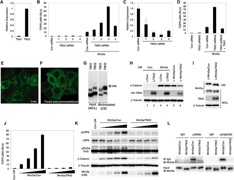
Figure 4. Tiki Inactivates Wnt3a(A) Relative expression levels of TIKI1 and TIKI2 mRNAs in HEK293T cells.(B) Knocking down endogenous TIKI2 in HEK293T cells enhanced Wnt3a signaling.(C) TIKI2 mRNA knockdown examined 48 hr after siRNA transfection.(D) The TIKI2 knockdown effect was countered by TIKI1 expression.(E and F) Immunofluorescence of HA∗Tiki1 expressed in HeLa cells. Live cells (E) or fixed-permeabilized cells (F) were labeled by an anti-HA antibody and a fluorescent secondary antibody.(G) HeLa cells expressing HA-TIKI1 or HA-TIKI2 were treated with a nonpermeable biotinylation reagent. Cell surface (CS) proteins were precipitated with streptavidin beads and blotted for HA-TIKI1/2. WCL: input whole-cell lysates. The slower-migrating form of HA-TIKI, due possibly to glycosylation, was enriched on the cell surface.(H) TIKI2 inhibited β-catenin stabilization in Wnt3a-expressing cells. Indicated L cell lines were also treated with control or Wnt3a CM for 2 hr (lanes 1–4). β-tubulin: a loading control.(I) Wnt3a was secreted similarly with or without TIKI2 but exhibited faster migration due to TIKI2.(J) Wnt3a CM from TIKI2-expressing L cells exhibited minimal activity, tested at concentrations in 2-fold dilutions.(K) Wnt3a CM from TIKI2-expressing L cells induced minimal LRP6 or Dvl2 phosphorylation or β-catenin stabilization at 2 hr. LRP6 intensity reduction was not due to decreased protein levels but due to LRP6 phosphorylation, which perturbed recognition by the LRP6 antibody used (not shown).(L) Wnt3a secreted from TIKI2-expressing cells exhibited minimal binding to mFz8CRD-IgG or LRP6N-IgG (Tamai et al., 2000).All error bars represent standard deviation (SD) in triplicates. See also Figure S4. Image published in: Zhang X et al. (2012) Copyright © 2012. Image reproduced with permission of the Publisher, Elsevier B. V. Larger Image Printer Friendly View |