XB-IMG-78646
Xenbase Image ID: 78646
|
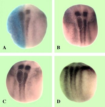
Fig. 5. RA signaling modifies XMeis3 expression along the A – P axis. Two-cell albino embryos were injected unilaterally into the animal hemisphere with XCYP26 or RALDH2 encoding RNAs. The injected side is on the left as marked by h-gal staining (blue) and all embryos are viewed dorsally with anterior on top and posterior at the bottom. (A) 0.5 ng of XCYP26 RNA, XMeis3 expression is shifted posteriorly in the hindbrain, and inhibited in the spinal cord in 100% of the embryos (86/86). The gap between hindbrain and spinal cord expression was narrowed or lost in 82% of the embryos (70/86). (B) 2.0 ng of RALDH2 RNA, XMeis3 expression is slightly shifted anteriorly, laterally expanded in the anterior spinal cord (as indicated with the dashed line) in 68% of the embryos (37/54). Expression in the hindbrain was not increased. (C) 2.0 ng of RALDH2 RNA and treatment with 500 nm all-trans-retinal (ATR) at stage 10.5. Similar phenotypes described in B were observed, including no increase in hindbrain expression, a slight anterior shift, and a lateral expansion of XMeis3 expression in 74% (35/48) of the embryos. (D) 18 nM RA treatment at gastrula stage 10.5. XMeis3 expression was strongly shifted anteriorly and increased in the spinal cord in 100% (76/76) of the embryos. Image published in: Dibner C et al. (2004) Copyright © 2004. Image reproduced with permission of the Publisher, Elsevier B. V. Larger Image Printer Friendly View |