Click here to close
Hello! We notice that you are using Internet Explorer, which is not supported by Xenbase and may cause the site to display incorrectly.
We suggest using a current version of Chrome,
FireFox, or Safari.
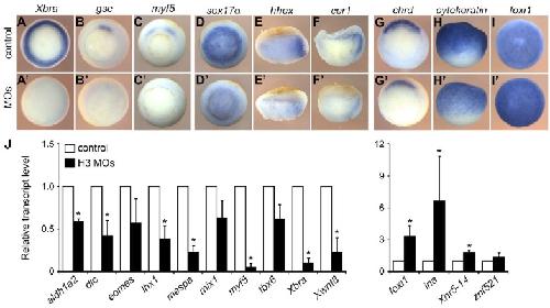
Fig. 2. H3.3/H3-depleted Xenopus embryos fail to express mesodermal marker genes. (A-Iâ²) Control and injected embryos fixed at early gastrula were subjected to RNA in situ hybridization for the analysis of mesodermal (A-Câ²), endodermal (D-Fâ²) and ectodermal (G-Iâ²) marker gene expression. Representative embryos from three experiments are shown. (A-D,G) Vegetal views; (E,F) lateral views of bisected embryos, dorsal towards the right; (H) lateral view; (I) animal view. (J) Expression of selected genes in control and H3 MO-injected embryos at stage 10.5 was measured by qRT-PCR. All values were normalized to ornithine decarboxylase (ODC) and plotted relative to the respective transcript levels in control embryos. Error bars indicate s.d. of three independent experiments. *P<0.05 using a two-tailed Student t-test.