XB-IMG-80461
Xenbase Image ID: 80461
|
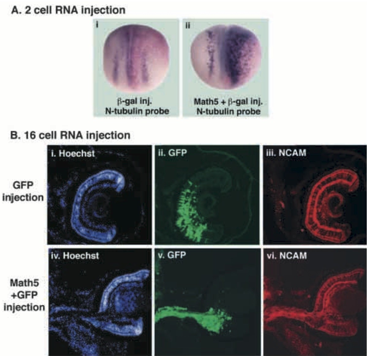
Fig. 5. Overexpression of Math5 in Xenopus embryos by RNA injection. (A) Injection into one cell of a 2-cell Xenopus embryo with RNA for nβgal alone (i) or in combination with Math5 (ii). Stage 14-15 embryos were stained to detect β-galactosidase activity (magenta) and probed by whole-mount in situ hybridization for N- tubulin expression (purple). Embryos are oriented in a dorsal view with anterior at the top and injected side on the right. (i) Control embryo expressing nβgal alone demonstrating the normal pattern of N-tubulin expression in the neural plate. (ii) Embryo expressing both Math5 and nβgal with ectopic N-tubulin on the injected side.
(B) Injection into blastomere D.1.1 of a 16-cell Xenopus embryo with RNA for GFP alone (i-iii) or with RNA for Math5 plus GFP (iv- vi). The embryos were fixed and cryostat sectioned at stage 41. Sections were immunostained for NCAM and labeled with Hoechst to visualize the retinal cell layers. (i-iii) Retinal section from a control embryo injected with GFP RNA alone. Hoechst staining reveals normal lamination (i). Within the retina, a large cluster of cells derived from the injected blastomere is labeled by GFP (ii) and robust NCAM staining is observed (iii). (iv-vi) A retinal section from an embryo injected with GFP and Math5 RNAs. Hoechst staining highlights a disruption in the normal arrangement of retinal cell layers (iv). This disrupted region corresponds to a cluster of GFP- labeled cells within the retina (v). The GFP-positive cells, including those extruding from the back of the retina, are NCAM-positive (vi). Scale bar 1 μm. Image published in: Brown NL et al. (1998) Copyright © 1998. Image reproduced with permission of the publisher and the copyright holder. This is an Open Access article distributed under the terms of the Creative Commons Attribution License.
Image source: Published Larger Image Printer Friendly View |