XB-IMG-80529
Xenbase Image ID: 80529
|
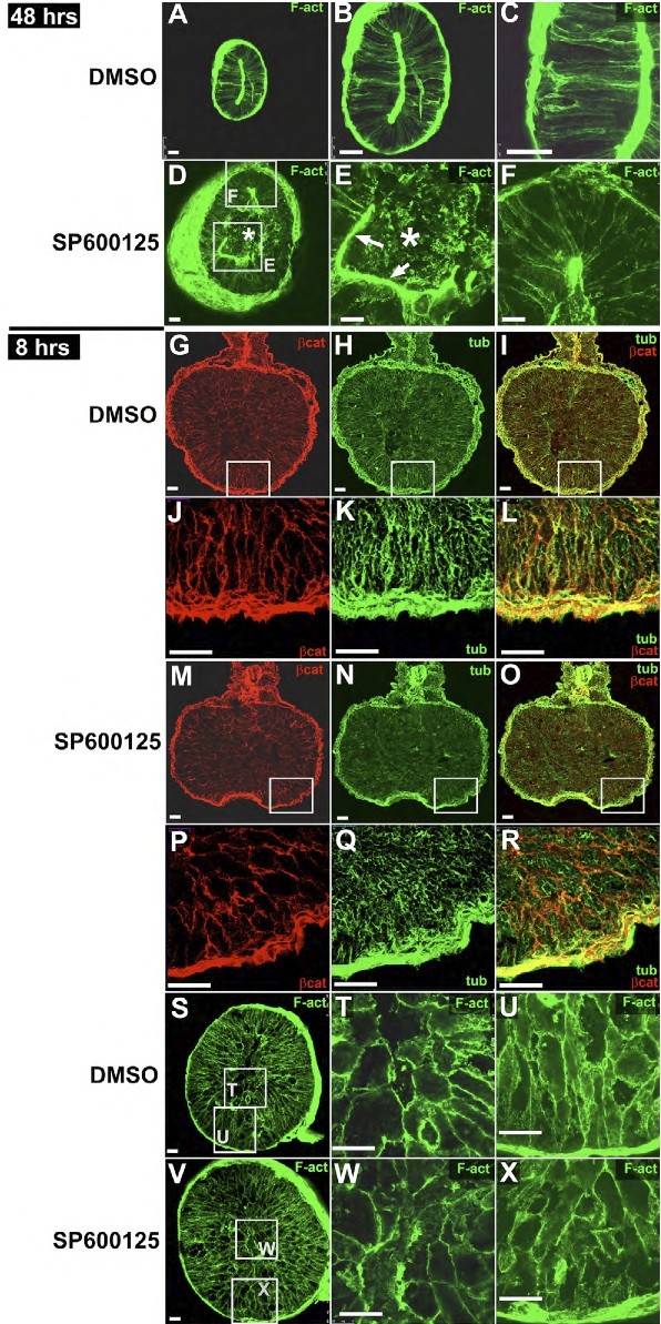
Fig. S6. JNK does not regulate actin polymerization in the gut. (A-F) Embryos were exposed to DMSO (A-C) or SP600125 (D-F) from stage 35 through stage 46 (48 hours). Whole gut tubes were bisected and stained with phalloidin to visualize the distribution of polymerized F-actin. E and F are higher magnifications of the boxed areas in D. Although actin is disrupted in the inner cell population (asterisks in D,E), cortical actin is still evident in the outer cells (F), and a robust actin belt forms at the apical surface of the abnormal epithelium (arrows, E). (G-X) Embryos were exposed to DMSO (G-L,S-U) or SP600125 (M-R,V-X) for 8 hours. Transverse sections of a representative embryo exposed to DMSO (G-L) or SP600125 (M-R) were stained to reveal the presence of α-tubulin (green; MTs) or β-catenin (red; adhesion), as indicated, or whole gut tubes were bisected and stained with phalloidin to visualize F-actin (S-X). Although the 8-hour exposure to SP600125 has disrupted the parallel bundling of MT arrays and the regular distribution of β-catenin at the membrane, there is no observable difference in cortical actin distribution in either the inner (W) or outer (X) endoderm cells. (J-L,P-R) Higher magnification images of the boxed regions in G-I,M-O, respectively. (T-U,W-X) Higher magnifications of the boxed areas in S and V, respectively. Scale bars: 50 μm. Image published in: Dush MK and Nascone-Yoder NM (2013) Copyright © 2013. Image reproduced with permission of the publisher and the copyright holder. This is an Open Access article distributed under the terms of the Creative Commons Attribution License. Larger Image Printer Friendly View |