XB-IMG-81002
Xenbase Image ID: 81002
|
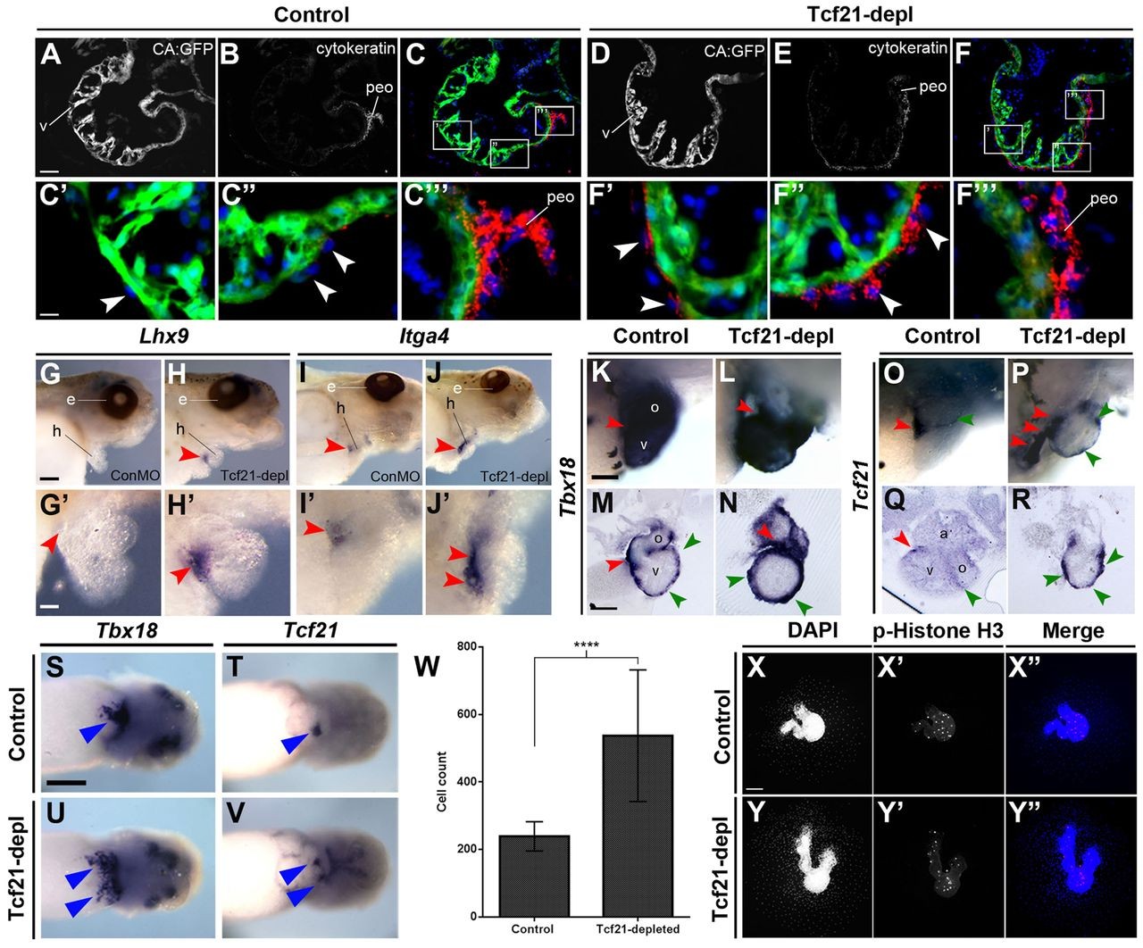
Fig. 7.
Tcf21 is required for correct specification of precursor PE cells and for epicardial maturation. (A-F‴) Transverse sections through the cardiac region of stage 46 CA:GFP transgenic Xenopus embryos stained for cytokeratin (red) and with DAPI (blue), with the myocardium expressing GFP under the cardiac actin promoter. Punctate cytokeratin staining is observed in migrating PE cells (arrowheads). (G-J′) ISH of stage 46 embryos showing the PEO markers (red arrowheads) Lhx9 and Itga4; lateral views with head facing right. G′-J′ are magnifications from G-J. (K-R) ISH of Tbx18 and Tcf21 in stage 46 embryos, showing lateral magnified views of hearts (K,L,O,P; anterior right) and transverse gelatin vibratome sections (M,N,Q,R; dorsal to top). Red arrowheads indicate PEO expression, green arrowheads migrating epicardial cell expression. Note the thickened and more rounded appearance of the Tbx18-expressing layer in Tcf21-depleted embryos (N). (S-V) ISH of Tbx18 and Tcf21; ventral images of younger, stage 40 embryos (anterior right). Arrowheads indicate PEO expression and duplication/expansion thereof in Tcf21-depleted embryos (U,V). (W) The number of migrating epicardial cells from Tcf21-depleted cardiac explants (17 control hearts, 23 Tcf21-depleted hearts, two independent experiments) is significantly increased compared with control (****P<0.0001, two-tailed unpaired non-parametric Mann-Whitney test). Mean s.d. (X-Y″) The increased number of DAPI-stained nuclei in Tcf21-depleted explants, compared with controls, is not due to an increase in proliferation as shown by the absence of phospho-Histone H3 staining. a, atrium; oft, outflow tract; peo, proepicardium; v, ventricle. Scale bars: 50 μm in A; 10 μm in C′; 500 μm in K; 100 μm in M; 2 mm in G,S; 1 mm in G′,X. Image published in: Tandon P et al. (2013) Copyright © 2013. Image reproduced with permission of the publisher and the copyright holder. This is an Open Access article distributed under the terms of the Creative Commons Attribution License. Larger Image Printer Friendly View |