XB-IMG-81357
Xenbase Image ID: 81357
|
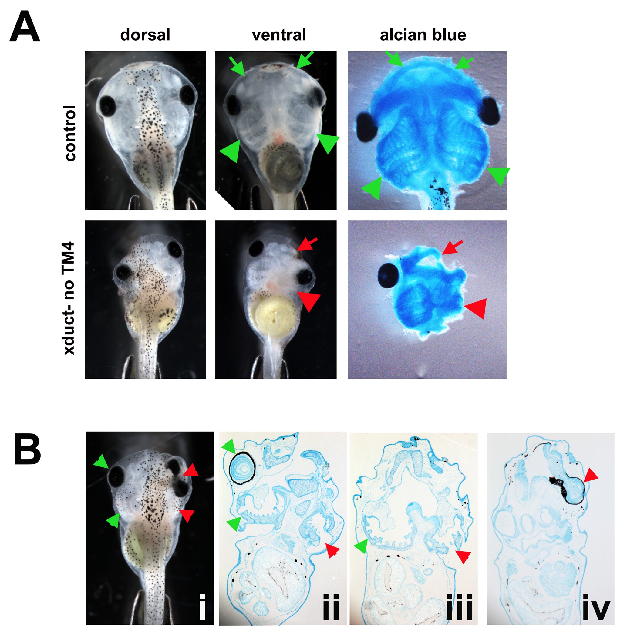
Supporting Figure 1 Alcian blue staining indicates that ductin inhibition affects facial cartilage morphology at stage 45. A: Embryos injected with xduct-noTM4 and uninjected embryos were fixed, stained with alcian blue, and the soft tissues were dissected away from the cartilage. Arrowheads indicate branchial arches, arrows indicate jaws; in all images, red indicates abnormal and green indicates normal morphologies. Alcian blue staining clearly indicates that both the jaw and branchial arch cartilage were affected by ductin inhibition. B: An embryo injected with xduct-noTM4 was imaged at stage 45 (B.i), fixed, stained with alcian blue, embedded in paraffin, and sectioned. B.ii: Ventral sections show a normal-sized and shaped eye on the left side, and a small branchial arch on the right side. B.iii: More dorsal sections also show a small branchial arch on the right side. B.iv: The right eye is actually two fused eyes, which are joined to the olfactory epithelium. This is also apparent in the image of the whole animal (B.i) Image published in: Vandenberg LN et al. (2011) Copyright © 2011. Image reproduced with permission of the Publisher, John Wiley & Sons.
Larger Image Printer Friendly View |