XB-IMG-81809
Xenbase Image ID: 81809
|
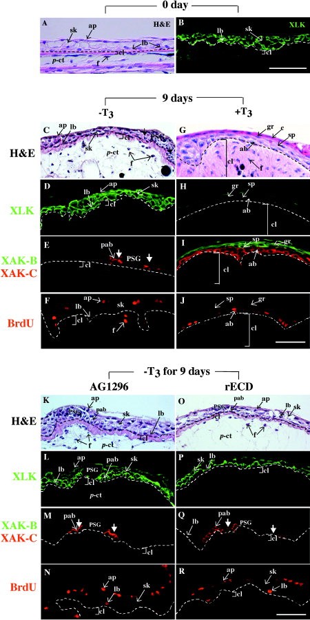
Fig. 3. Culture of larval skin in the presence
of thyroid hormone (TH) or inhibitors
of platelet-derived growth factor (PDGF)
signaling. Pieces of back skin were isolated
from tadpoles at stage 54/55 (A,B)
and were cultured for 9 days in the absence
(-T3; C) or presence of ( T3; G)
100 nM 3,3 ,5-triiodothyronine (T3). A 10 M
AG1296 (K) or 100 g/ml recombinant
extracellular domain of PDGF receptor-
(rECD; O) was added for 9 days in the
absence of T3. AG1296 was dissolved in
0.1% dimethyl sulfoxide. A,C,G,K,O: Hematoxylin
and eosin (H&E) stains. B,D,H,L,P: Immunostaining
using anti-XLK antisera.
E,I,M,Q: Double immunochemical staining
using antiAK-C (shown in red) and XAK-B
(green) antisera. F,J,N,R: Skins were labeled
with 20 M bromodeoxyuridine
(BrdU) for the last 24 hr and were immunostained
by using anti-BrdU antibodies.
Dashed lines in the photographs represent
the location of the basement membrane.
Arrows with closed arrowheads in E, M, and
Q show XAK-Cositive preadult basal cells
around primordial secretory glands (PSG).
sp, spinous cells; gr, granular cells; c, cornified
cells. For other abbreviations, see
legend to Figure 2. Scale bars 50 m in B
(applies to A,B), in J (applies to C), in R
(applies to K). Image published in: Utoh R et al. (2003) Copyright © 2003. Image reproduced with permission of the Publisher, John Wiley & Sons.
Image source: Published Larger Image Printer Friendly View |