Click here to close Hello! We notice that you are using Internet Explorer, which is not supported by Xenbase and may cause the site to display incorrectly. We suggest using a current version of Chrome, FireFox, or Safari.
XB-IMG-82056

Xenbase Image ID: 82056

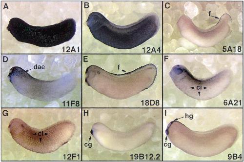
Fig. 6. Epidermal regionalization. Whole-mount in situ hybridizations of tailbud embryos are shown in lateral view. (A,B) Show pan-epidermal markers and (C) show genes with expression in various epidermal regions. cg, Cement gland; ci, ciliated cells; dae, dorsoanterior epidermis; f, fin; hg, hatching gland.

Image published in: Gawantka V et al. (1998)

Copyright © 1998. Image reproduced with permission of the Publisher, Elsevier B. V.

GeneSynonymsSpeciesStage(s)Tissue
krt12.4.Sck81, cytok, cytokeratin, E-keratin, epi-keratin, epidermal keratin, Epidermis-specific keratin, EpiK, epiker, EpK, epK, K81, xK81, xk81a, xk81a1X. laevisThroughout NF stage 29 and 30epidermis
msx2.LHhox 7.1X. laevisThroughout NF stage 29 and 30 
odad2.Sarmc4X. laevisThroughout NF stage 29 and 30ciliated epidermal cell
gale.LxgaleX. laevisThroughout NF stage 29 and 30cement gland
upk3a.Lup3a, upiii, upiiia, upk3, uroplakin III, xUPIIIX. laevisThroughout NF stage 29 and 30fin
atp6v0c.Latp6c, atp6l, atpl, ductin, vatl, vma3, xductX. laevisThroughout NF stage 29 and 30ciliated epidermal cell
anxa5.Lannexin A5, anx5, enx2X. laevisThroughout NF stage 29 and 30cement gland
hatching gland

Image source: Published

Larger Image
Printer Friendly View

Return to previous page