XB-IMG-82109
Xenbase Image ID: 82109
|
|
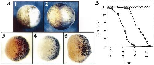
Fig. 4. Embryonic death is rescued by ectopic expression of Bcl-2 protein or injection of the caspase inhibitor z-DEVD-fmk into embryos. (A) Panels 1 and 2
show embryonic morphology, panels 3, 4 and 5 show TUNEL stained embryos. Embryos in panels 1 and 3 were irradiated at St. 1, 40 Gy, and 1 blastomere
was injected with Bcl-2 RNA at the 2-cell stage. Panel 1 shows a St. 10.5 embryo, cells in the left half of the embryo, i.e. those arising from the blastomere
injected with Bcl-2 RNA, look normal while the right half of the embryo is dying. In this experiment 24/28 embryos showed this phenotype and the
experiment is representative of three different experiments. Panel 3 shows a similarly treated embryo which was fixed and TUNEL stained at St. 10.5. Panel 2
shows a St. 10.5 embryo in which one blastomere was injected with z-DEVD-fmk at the 2 cell stage, to yield a final concentration of 00 mM in the embryo,
the left half of the embryo containing z-DEVD-fmk is protected from embryonic death. In this experiment 14/25 injected embryos showed this phenotype and
the result is representative of three different experiments. Panel 4 shows an embryo treated with a-amanitin at the 2-cell stage in which one blastomere was
injected with Bcl-2. The embryo was fixed and TUNEL stained at St. 10.5, no TUNEL staining is observed in the cells arising from the blastomere injected
with Bcl-2, left side of embryo. Panel 5 shows an embryo injected with Bcl-2 RNA at the 2-cell stage, followed by cycloheximide treatment at St. 7. This
allowed a period of h in which Bcl-2 protein could be synthesized. The embryo was fixed and TUNEL stained at a time equivalent to St. 10.5, and the cells
arising from the injected blastomere, left half of embryo, show no TUNEL staining. (B) Percent survival curve for irradiated embryos, +/- Bcl-2. Embryos
were irradiated (40 Gy) at the one cell stage, and both blastomeres injected with Bcl-2 RNA at the 2-cell stage. Percent survival refers to the % of surviving
embryos in a population, i.e. embryos that do not turn white, or embryos that do not have greater than 50% of the cell on their surface appearing white.
Control embryos (W), g-irradiated embryos (B), g-irradiated embryos + Bcl-2 (X). Percentages were scored on batches of 50 embryos, and the data are
representative of three separate experiments. Image published in: Hensey C and Gautier J (1997) Copyright © 1997. Image reproduced with permission of the Publisher, Elsevier B. V. Larger Image Printer Friendly View |