XB-IMG-82243
Xenbase Image ID: 82243
|
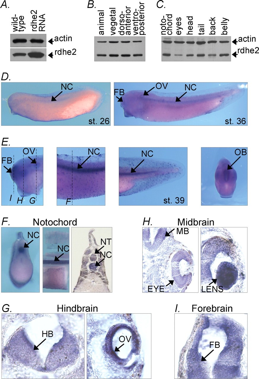
FIGURE 4. Expression pattern of endogenous rdhe2 in frog embryo. A, Western blot analysis of extracts (50 μg of total protein) of neurula stage wild-type embryos or embryos injected with in vitro synthesized rdhe2 mRNA revealing a single major band of the expected size (∼34 kDa), which is increased in injected embryos, thus confirming that antibody is specific. B and C, Western blot analysis of dissected gastrula embryos (B) and stage 39 tadpoles (C), 30 μg of total protein per lane. D, whole mount in situ hybridization with antisense rdhe2 probe. E, in situ hybridization of dissected embryos with rdhe2 probe. Dashed lines indicate where embryos were cut in F. F, embryos stained with rdhe2 probe further dissected to better reveal staining of notochord (left, cross-section through the trunk; center, sagittal cut through the upper portion of the trunk), or paraffin-embedded and sectioned at 50 μm (right). G, 50-μm sections of prestained paraffin-embedded specimen revealing staining in the brain, eye, and otic vesicle. NC, notochord; NT, neural tube; FB, forebrain; MB, midbrain; HB, hindbrain, OV, otic vesicle; OB, olfactory bulb. Image published in: Belyaeva OV et al. (2012) Copyright © 2012. Image reproduced with permission of the publisher and the copyright holder. This is an Open Access article distributed under the terms of the Creative Commons Attribution License.
Image source: Published Larger Image Printer Friendly View |