XB-IMG-82517
Xenbase Image ID: 82517
|
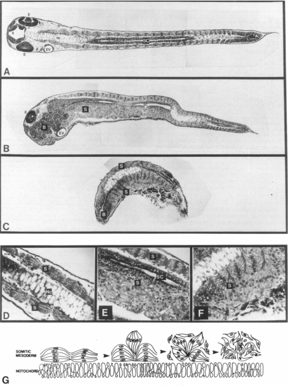
Figure 4. Histological Examination of Normal and Xhox-IA Injected Embryos at the Tailbud Stage (A) Horizontal section of a normal embryo at the level of the notochord and spinal chord. (8) Horizontal section of a Xhox-IA injected embryo at the level of the spinal chord and brain. (C) Horizontal section of a severely kinked and stunted Xhox-IA injected embryo at the level of the brain and notochord. (0) A 2.5x enlargement of a part of the normal embryo shown in (A). (E) A 2.5x enlargement of a part of the Xhox-IA injected embryo in (8) showing the loss of somite units in the lower or injected side. (F) A 2.5x enlargement of a part of the Xhox-1A injected embryo in (C) showing the somite-like units interdigitated between the notochord-proximal somites on the lower or injected side (see text). (G) Diagram of the differences in the severity of the Xhox-1A phenotypes from normal on the left to totally chaotic on the right. B is the brain, E is the eye, EV is the ear vesicle, NC is the notochord, S is the somitic mesoderm, and SC IS the spinal cord. Image published in: Harvey RP and Melton DA (1988) Copyright © 1988. Image reproduced with permission of the Publisher, Elsevier B. V. Larger Image Printer Friendly View |