XB-IMG-82518
Xenbase Image ID: 82518
|
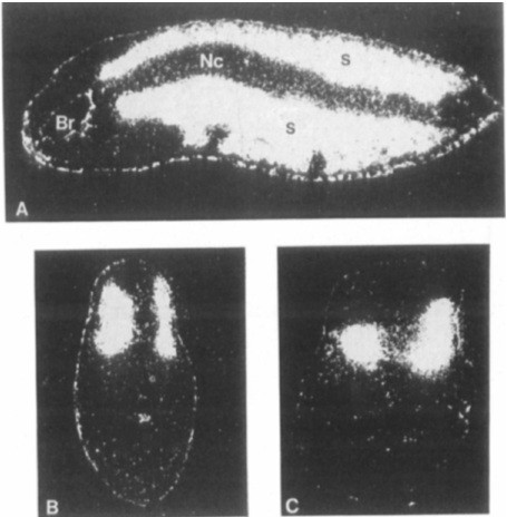
Figure 5. In Situ Hybridization to Xhox-IA Injected Embryos Using a Muscle-Specific a-Actin Probe (A) Horizontal sectron of Xhox-1A embryos viewed with dark field op- tics. Hybridization grains are seen as white. The embryo is outlined in pigment granules which also appear as white grains in these photo- graphs The normal or uninjected side of the embryo is uppermost. Note the two patches of hybridization-negative cells on the lower side of the disturbed tissue. Br is the brain, NC is the notochord, and S is the somitic mesoderm. (8) and (C). Transverse sections of Xhox-IA in- jected embryos viewed with dark field optics. Disturbed tissue is on the left side of each case. Note the asymmetry in the shape of each pair. Image published in: Harvey RP and Melton DA (1988) Copyright © 1988. Image reproduced with permission of the Publisher, Elsevier B. V. Larger Image Printer Friendly View |