XB-IMG-82878
Xenbase Image ID: 82878
|
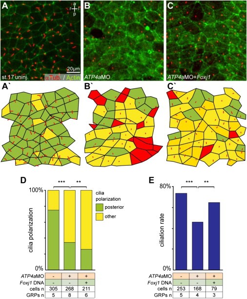
Figure S4. Rescue of Ciliation Rate but Not Cilia Polarization by Foxj1 in ATP4a Morphants, Related to Figure 5Embryos were injected at the 4-cell stage into the DMZ and dorsal explants were prepared at stage 17. Specimens were processed for IHC to assess cilia polarization (A-D), or for SEM analysis to determine the GRP ciliation rate (E).(A�C) IHC using antibodies against acetylated tubulin to visualize cilia (red) and actin (green) to outline cell boundaries. (A) Control uninjected specimen. (B) ATP4a morphant. (C) Coinjection of ATP4aMo and Foxj1 mRNA. (Aâ²-Câ²) Evaluation of results.(D) Quantification of cilia polarization. Note that co-injection of Foxj1 aggravated polarization defects.(E) Ciliation rate. Note that ciliation rate was partially rescued by Foxj1 co-injection. Image published in: Walentek P et al. (2012) Copyright © 2012. Image reproduced with permission of the Publisher and the copyright holder. This is an Open Access article distributed under the terms of the Creative Commons Attribution License. Larger Image Printer Friendly View |