XB-IMG-82997
Xenbase Image ID: 82997
|
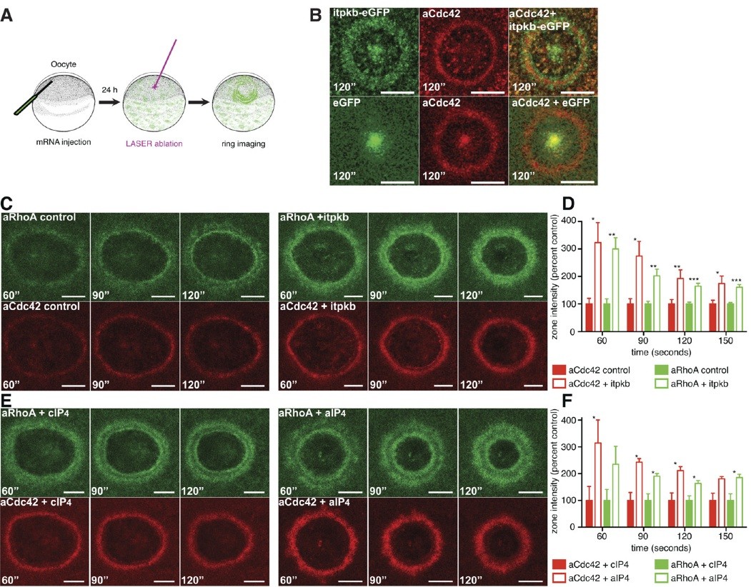
Fig. S5. Related to Fig. 4. Itpkb and InsP4 increase wound-induced activation of RhoA and Cdc42 in single-cell wounds. (A) Schematic diagram showing the oocyte assay in analyzing the activity of Cdc42 and RhoA after single-cell wounding. (B) Itpkb accumulates around wound edge. (Upper) An oocyte injected with 5 ng itpkb-eGFP mRNA and activated Cdc42 (aCdc42) probe. (Lower) A control oocyte injected with 1.5 ng eGFP mRNA and aCdc42 probe. itpkb-eGFP n = 7, eGFP n = 9. (C) Itpkb increases the levels of active Cdc42 and active RhoA. (Left) A wounded control oocyte injected with aRhoA (green) and aCdc42 (red) probes. (Right) A wounded oocyte injected with both aRhoA and aCdc42 probes and 1 ng myc-itpkb mRNA. (D) Quantification of aRhoA and aCdc42 zone intensities over time in the presence of myc-itpkb, relative to controls (probes alone). Control n = 11, itpkb n = 7. (E) Ins(1,3,4,5)P4 increases the levels of active Cdc42 and active RhoA. (Left) A wounded control oocyte injected with aRhoA and aCdc42 probes and incubated with 0.5 μM cIP4 (IP4 control). (Right) Wounded oocytes injected with both probes and incubated with 0.5 μM aIP4 (IP4 active). (F) Quantification of aRhoA and aCdc42 zone intensities over time in the presence of 0.5 μM aIP4, relative to controls (0.5 μM cIP4). cIP4 n = 5, aIP4 n = 5. Each time point was analyzed by Student t test, nonparametric Mann-Whitney test. Results are shown as means � SEM. *P < 0.05, **P < 0.01, ***P < 0.001. (Bars, 20 μm.) Image published in: Soto X et al. (2013) Copyright © 2013. Image reproduced with permission of the publisher and the copyright holder. This is an Open Access article distributed under the terms of the Creative Commons Attribution License. Larger Image Printer Friendly View |