XB-IMG-83617
Xenbase Image ID: 83617
|
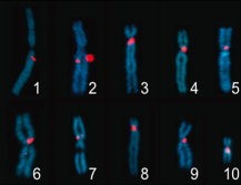
Figure 2. Cytological localization of centromeric markers. Fluorescence in situ hybridization (FISH) probes were generated from cDNAs of genes on centromere-linked scaffolds identified by gynogenesis. Probe name, location: Chr1/LG1: mast3, q0.13; Chr2/LG6: epb41, q0.22; Chr3/LG8: gemin5, p0.00; Chr4/LG7: znf423, p0.00; Chr5/LG9: olig3, p0.00; Chr6/LG2: fbxl7, q0.13; Chr7/LG4: mat1a, p0.12; Chr8/LG5: naif1, p0.00; Chr9/LG3: stat4, q0.09; Chr10/LG10: ezh1, q0.03.Download figure to PowerPoint Image published in: Khokha MK et al. (2009) Copyright © 2009. Image reproduced with permission of the Publisher, John Wiley & Sons. Larger Image Printer Friendly View |