XB-IMG-83754
Xenbase Image ID: 83754
|
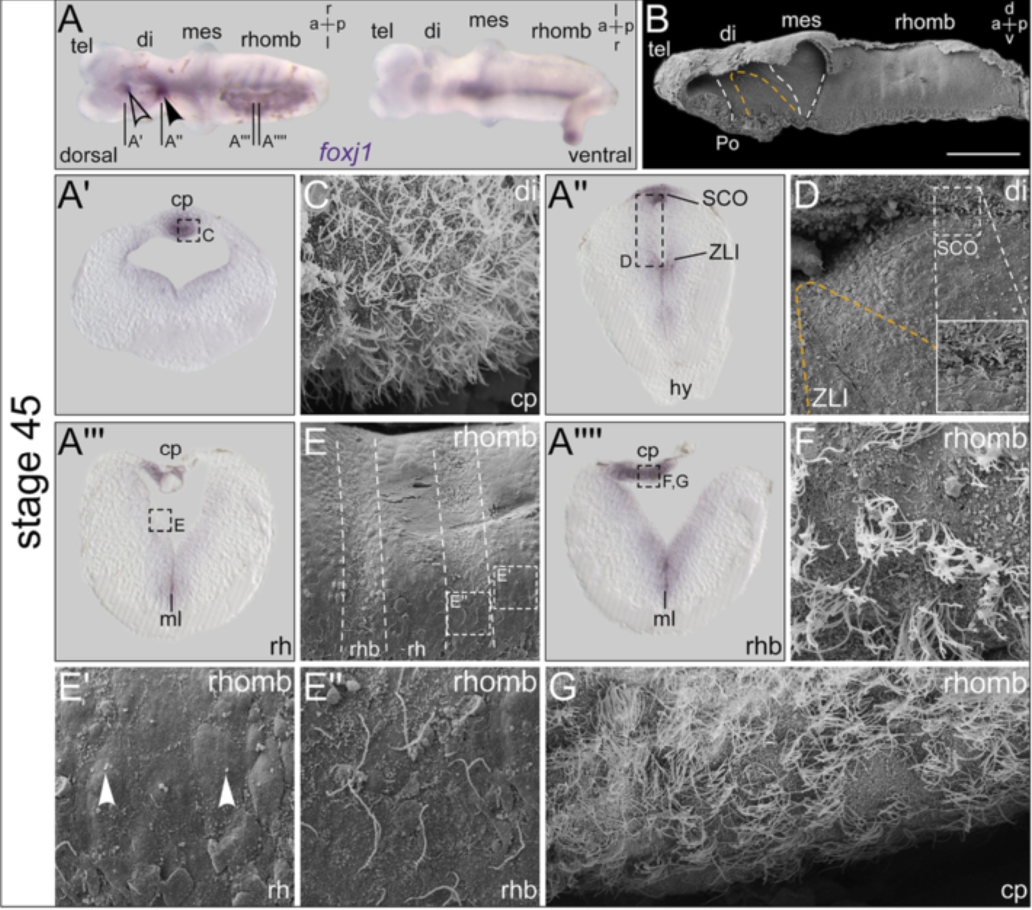
Figure 3. Additional foxj1 expression domains identify regions with emerging ciliation. In situ hybridization and scanning electron microscopy (SEM) on explanted brains at stage 45. (A) Explant shown in dorsal (d) and ventral (v) view. Expression in the ventral midline, rhombomere boundaries, subcommissural organ (SCO, arrowhead), and the choroid plexus (cp; outlined arrowhead). (B) SEM picture of brain explant dissected sagittally with view onto the ventricular surface, the zona limitans intrathalamica (ZLI) is delimited with orange dashed line and boundaries between brain regions are indicated by white dashed lines. Bar represents 200 ?m. (A?-A??) Transversal histological sections as indicated in (A), dorsal side up, and SEM pictures (C-G) of corresponding regions as indicated in the sections, respectively. (C) MCCs on cp invaginating from the diencephalon roof. (D) Close-up view onto the ZLI region; enlargement showing ciliated structure of the SCO. (E) Close-up view revealing metamerical organization of the rhombencephalon (rhomb). Rhombomeres (rh) and rh boundaries (rhb) indicated by dashed lines. (E?) Enlargement of the rh region; arrowheads pointing to short primary cilia. (E?) Enlargement of the rhb with cells bearing elongated monocilia. (F, G) Close-up views onto the fourth ventricle cp showing MCCs. a = anterior; di = diencephalon; hy = hypophysis; l = left; mes = mesencephalon; ml = midline; p = posterior; Po = preoptic region; r = right; tel = telencephalon. Image published in: Hagenlocher C et al. (2013) Copyright © 2013 Hagenlocher et al. Creative Commons Attribution license Larger Image Printer Friendly View |