XB-IMG-83761
Xenbase Image ID: 83761

|
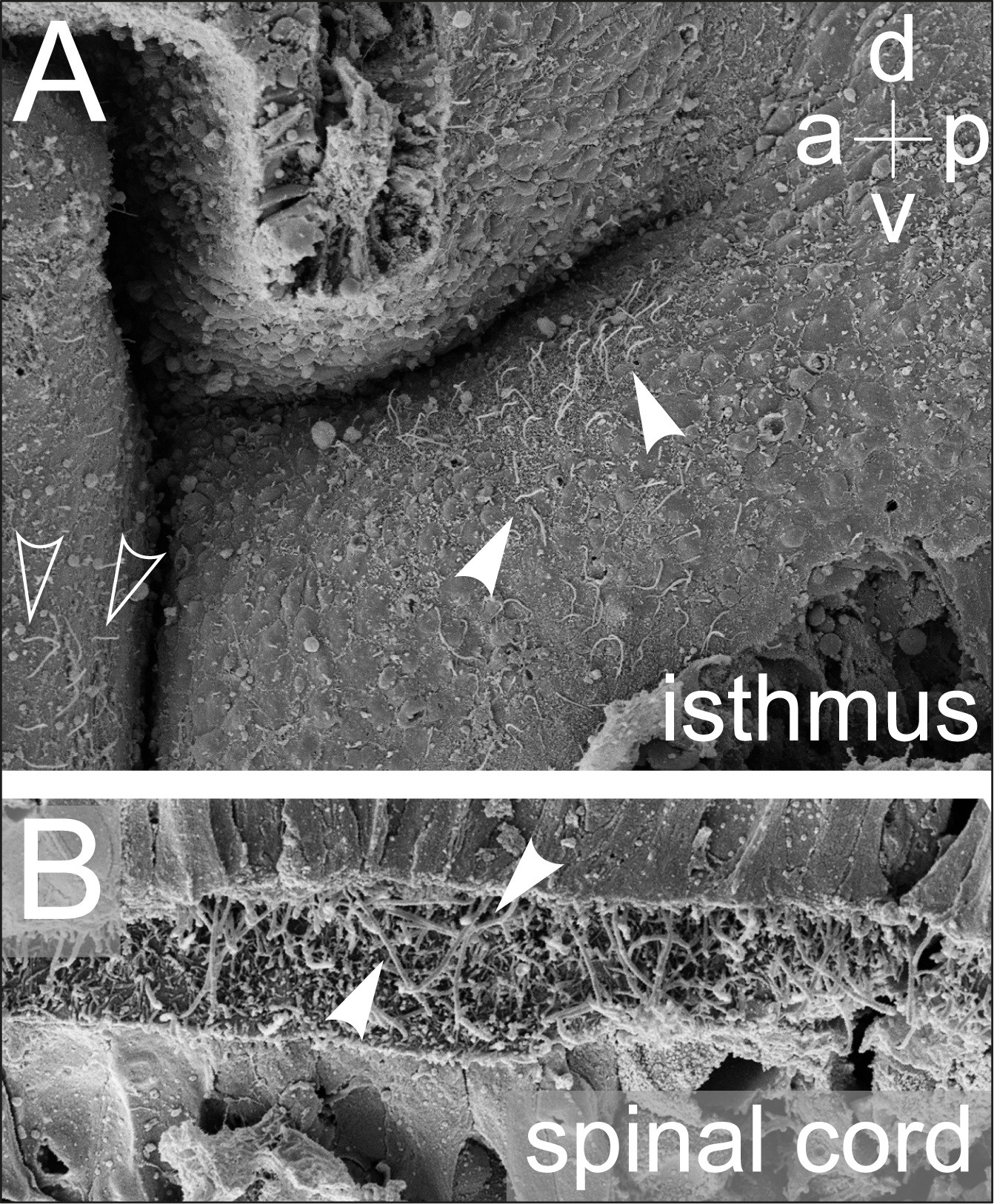
Figure S4. Signaling centers in the central nervous system show elongated monocilia. Scanning electron microscopy pictures of sagittally bisected brain explants at stage 45. (A) The isthmus organizer (mid-hindbrain boundary) region; note the presence of several elongated monocilia on the mesencephalic aqueduct side (outlined arrowheads) as well as a population of monociliated cells on the ventral (v) aspect of the isthmus (arrowheads). (B) Close-up view onto the lumen of the spinal cord. Arrowheads point to elongated monocilia projecting into the central canal, the appearance of which correlates with expression of foxj1 (cf Figure 1B-F, Figure 2A). a = anterior; d = dorsal;
p = posterior. Image published in: Hagenlocher C et al. (2013) Copyright © 2013 Hagenlocher et al. Creative Commons Attribution license Larger Image Printer Friendly View |
