XB-IMG-84096
Xenbase Image ID: 84096
|
|
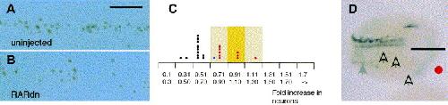
Fig. 3. Depressed retinoid signalling decreases the number of Isl1+ primary sensory neurons. (A,B). (A) Isl1 expression in the primary sensory neuron domain of a normal embryo. (B) Embryos injected with RARdn mRNA have a reduced number of Isl1+ primary sensory neurons. In this example the width of the domain is wider in the RARdn injected embryos than normal. Scale bar applies to (A) and (B) and is equal to 60 μm. (C) Analysis of the effect of unilaterally injecting the xRARα2dn mRNA on the ratio of Isl1+ primary sensory neurons on the injected compared to the uninjected sides of the embryo. The yellow background indicates the peak and distribution range of Isl-1+ primary sensory neurons in normal embryos (shown in Fig. 2D). Blue dots indicate embryos injected with 1.0 ng and red dots embryos injected with 0.5ng RARdn mRNA. In contrast to Fig. 2D there is now a shift towards reduced numbers of Isl1+ primary sensory neurons on the injected side of the embryo. (D) NST expression assayed by wholemount in-situ hybridization in an embryo unilaterally injected with RARdn mRNA. The injected side is marked with a red dot and the midline by a bar. The expression of NST in the motor, and inter neuron domains is undetectable, and is also missing from the trigeminal ganglion domain (open arrows). Expression in the sensory neuron domain is detectable (grey arrow) but much weaker than on the control side of the embryo. Image published in: Sharpe C and Goldstone K (2000) Copyright © 2000. Image reproduced with permission of the Publisher, Elsevier B. V. Larger Image Printer Friendly View |