XB-IMG-85181
Xenbase Image ID: 85181
|
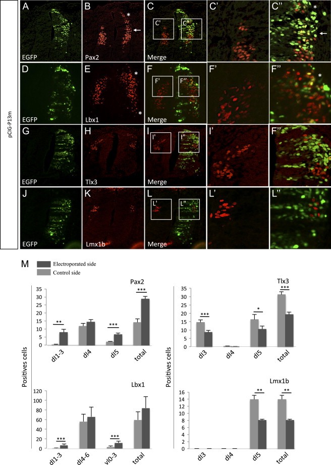
Fig. 9. Prdm13 overexpression induces GABAergic and suppresses glutamatergic markers in the chick neural tube. (A–L) Immunofluorescence on transverse sections of HH24–25 chick neural tube, with the right side of the neural tube electroporated with a mouse pCIG-Prdm13 expression construct with antibodies against Pax2, Lbx1, Tlx3, Lmx1b in red and GFP in green. Note the disappearance of the Pax2 gap of staining between the presumptive dI4 and dI6 neurons (arrow) and the Pax2 staining above dI4 (asterisk). Lbx1 is also detected more ventrally and dorsally (asterisks) upon Prdm13 overexpression. In contrast, Tlx3 in dI3/5 and Lmx1b in dI5 are decreased. High magnification views of the control and electroporated sides in the dorsal half of the neural tube are shown in C'–C'' and F'–F''. In contrast, in I'–I'' and L'–L'', no GFP+ Prdm13 expressing cells are Tlx3+ or Lmx1b+. (M) Quantification of the number of Pax2, Tlx3, Lbx1 and Lmx1b positive cells in the different progenitor regions on the Prdm13 electroporated side compared to the contralateral, control side. P<0.05*, P<0.01**, P<0.001***; (n≥3). Image published in: Hanotel J et al. (2014) Copyright © 2014. Image reproduced with permission of the Publisher, Elsevier B. V. Larger Image Printer Friendly View |