XB-IMG-86755
Xenbase Image ID: 86755
|
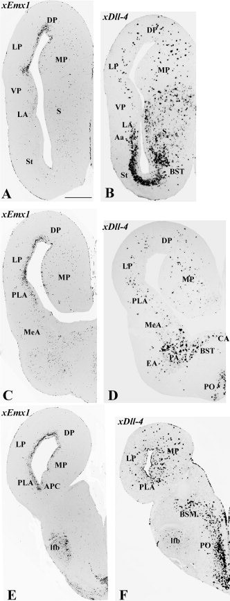
Figure 6. Frontal paraffin sections through the telencephalon of an adult Xenopus, from intermediate (top) to caudal (bottom) levels, hybridized for xEmx1 (A,C,E) or xDll4 (B,D,F). Note the strong xEmx1 expression in the majority of the pallium (specially in the dorsal and lateral divisions) except for the ventral pallium. Only a few xEmx1-expressing cells are observed in the VP mantle. In the pallium, including the ventral pallium, only some scattered mantle cells express the subpallial marker gene xDll4, and these are probably immigrant cells of subpallial origin. The caudal part of the lateral amygdalar nucleus, here called the posterolateral amygdalar nucleus (PLA), is rich in xEmx1 expression and may be a lateral pallial derivative. On the other hand, the medial amygdalar nucleus is poor in both xEmx1 and xDll4, and at least a major part of it may be a ventral pallial derivative. See text for more details. For abbreviations , see list. Scale bar = 200 μm in A (applies to all). Image published in: Brox A et al. (2004) Copyright © 2004. Image reproduced with permission of the Publisher, John Wiley & Sons.
Image source: Published Larger Image Printer Friendly View |