XB-IMG-86892
Xenbase Image ID: 86892
|
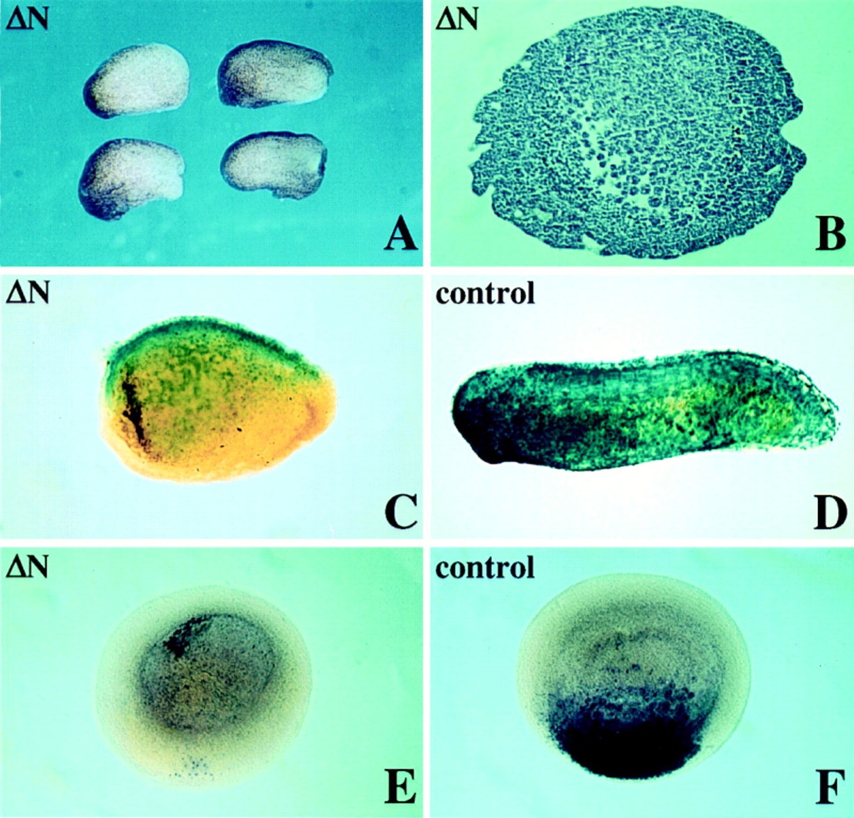
Figure 7.
Suppression of Endogenous Axis Formation
(A–D) Embryos allowed to develop until sibling stage 27. Anterior is to the left. (A) shows phenotypes of embryos injected with 250 pg of ΔN RNA in both dorsal blastomeres at the 4-cell stage. (B) shows a horizontal section of an embryo as in (A), with blastopore to the right.
(C and D) Embryos injected with a combination of 250 pg of ΔN RNA and 250 pg of β-gal RNA (C) or β-gal RNA alone (D).
(E and F) In situ hybridization with goosecoid on embryos injected with 250 pg of ΔN (E) or 250 pg of β-gal (F) analyzed at stage 11. Dorsal view of cleared embryos with the dorsal blastopore lip to the bottom. Some nonspecific staining occurs in the blastocoel cavity in (E). Image published in: Molenaar M et al. (1996) Copyright © 1996. Image reproduced with permission of the Publisher, Elsevier B. V. Larger Image Printer Friendly View |