| Images | Sources | Experiment + Assay | Phenotypes | Human Diseases | ||
|---|---|---|---|---|---|---|

|
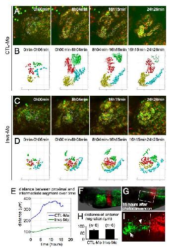
Fig. 2 C D E
Lienkamp S et al. (2010) |
Xla Wt + invs MO
NF29/30-40 (Other Detection Assay) |
|
nephronophthisis 2
|
||

|
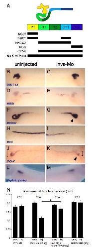
Fig. S2 B D F H J
Lienkamp S et al. (2010) |
Xla Wt + invs MO
NF33/34-40 (in situ hybridization) |
|
nephronophthisis 2
|
||

|
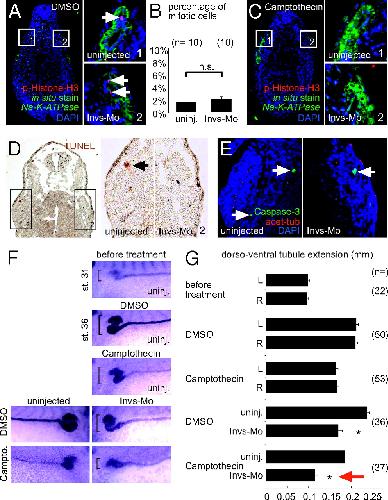
Fig. 3 F G
Lienkamp S et al. (2010) |
Xla Wt + invs MO
NF35/36 (in situ hybridization) |
|
nephronophthisis 2
|
||

|
Fig. S 4 E G
Lienkamp S et al. (2010) |
Xla Wt + invs MO
NF37/38 (in situ hybridization) |
|
nephronophthisis 2
|
||

|
Fig. S 4 K N
Lienkamp S et al. (2010) |
Xla Wt + invs MO
NF37/38 (in situ hybridization) |
|
nephronophthisis 2
|
||

|
Fig. 1 D F
Lienkamp S et al. (2010) |
Xla Wt + invs MO
NF40 (immunohistochemistry) |
|
nephronophthisis 2
|
||

|
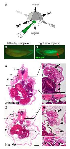
Fig. 1 G H J N
Lienkamp S et al. (2010) |
Xla Wt + invs MO
NF42 (Other Detection Assay) |
|
nephronophthisis 2
|
||

|
Fig. 1 B
Lienkamp S et al. (2010) |
Xla Wt + invs MO
NF43-45 (whole-mount microscopy) |
|
nephronophthisis 2
|
||

|
Fig. S1 G H
Lienkamp S et al. (2010) |
Xla Wt + invs MO
NF44 (histology) |
|
nephronophthisis 2
|