XenHead: cranial development
XenHead: Illustrations for Facial and Cranial Development
 The XenHead project, led by Drs. Dany. Adams and Mike Levin from Tufts University, commissioned new artwork to complement, and fill in some gaps, in the classic Xenopus development series by Nieuwkoop and Faber from 1967.
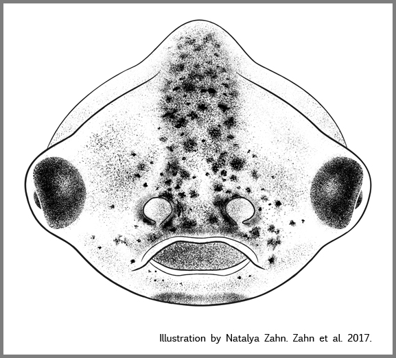
The resulting drawings by illustrator, Natalya Zahn, closely match the aesthetic of the familiar NF stage series, adding new anterior views of NF stages 22-50, when the cranio-facial features develop.
Importantly, the new illustrations are Open Access, distributed under the terms of the Creative Commons Attribution License (https://creativecommons.org/licenses/by-nc/4.0/), which permits unrestricted use, distribution and reproduction in any medium provided that the original work is properly attributed.
What this means to you:
• Free use of any/all of the Zahn drawings for non-commercial purposes, such as lecture slides, handouts, lab notebooks, lab worksheets, posters, power-point slides.
• Free use of up to 6 of the Zahn drawings per published article! The same image can be re-used multiple times in figures and diagrams, but you are limited to 6 unique images/article.
Want to use more than 7 images?
• Contact illustrator Natalya Zahn [natalya@natalya.com] for permission to use 7 or more illustrations (Fees may apply).
To use the Zahn drawings in your published work, do these 3 things:
1) Write this in captions of any figures: Xenopus illustrations by Natalya Zahn (2017).
2) Cite Xenbase as the source of the illustrations: Xenbase (www.xenbase.org RRID:SCR_003280), and
3) Cite this article: The Zahn drawings: new illustrations of Xenopus embryo and tadpole stages for studies of craniofacial development. Natalya Zahn, Michael Levin, Dany Spencer Adams. Development 2017. 144: 2708-2713; doi: 10.1242/dev.151308
Want to use the Zahn drawings in a commercial publication, like a text book or science magazine?
• Request permission from the Illustrator Natalya Zahn [natalya@natalya.com] to use any illustration in a commercial publication (Fees apply).
Access the original Zahn drawings article:
• Click here to view article at Development.
• Click here to view article on Xenbase.
Published in Development: Xenbase engaged with Natalya Zahn to create another 130+ drawings to provide a full set of open access illustrations of Xenopus development.
Like the classic N&F drawings, the updated Xenopus drawings by Natalya Zahn are a comphrehemsive development series including anterior, dorsal, ventral, and lateral views from early embryos and late tadpole stages, a progression of limb development, as well as a few ventral diagrams showing internal organ development.
Importantly, the entire series of Zahn drawings, for the XenHead and Xenbase commissions will remain Open Access and free to use for non-commercial purposes (limitations will still apply, namely a maximum of 6 illustrations per publication). Commercial use of the images will require payment of fees to the illustrator, Natalya Zahn.
The Zahn Drawings: downloadable from Xenbase
Click on the views below to see all available illustrations. Click any drawing to view larger image.
To download individual illustrations, mouse over and right-click, or ctrl-click, and choose "Save image as..." [and name the file], or choose "Copy image", the paste the file into imaging program.
Click here to download all of the simple line drawings (zip file).


Last Updated: 2019-09-01