Inner ear histology
Xenopus laevis inner ear histology
Viviane Wilms and Hans Gerd Nothwang
Neurogenetics group, Cluster of Excellence “Hearing4All”, School of Medicine and Health Sciences, Carl von Ossietzky University Oldenburg, 26111 Oldenburg, Germany
Histological methodology and captions (view pdf).
Images and methodology provided by Viviane Wilms.
[click image to view enlargement]
, Figure 2.")
.")
Adapted from Bever et al. (2003), Figure 2. Paint-filled inner ears (pseudo-colored green) are shown in intact, fixed Xenopus laevis specimens at stage 28 and stage 52 to demonstrate the location and relative size of the developing ear. Images are lateral views. Inner ears were filled with paint to visualize the inner surface of the membranous labyrinth. The endolymphatic sac is visible in the stage 52 specimen (arrow- head), even though it did not fill with paint. Total length of stage 52 specimen is 25 mm from tip of snout to tip of tail. Scale bar: 1.0 mm. Based on figure 2 of Bever et al. (2003).
Higher resolution figure.
Higher resolution figure.
Schematic of Xenopus laevis inner ear anatomy. Section markers. Anterior to the left, dorsal up (orientation as shown in figure above). Redrawn from Bever et al. (2003).
Higher resolution figure.
Higher resolution figure.
Tadpole: NF stage 52.


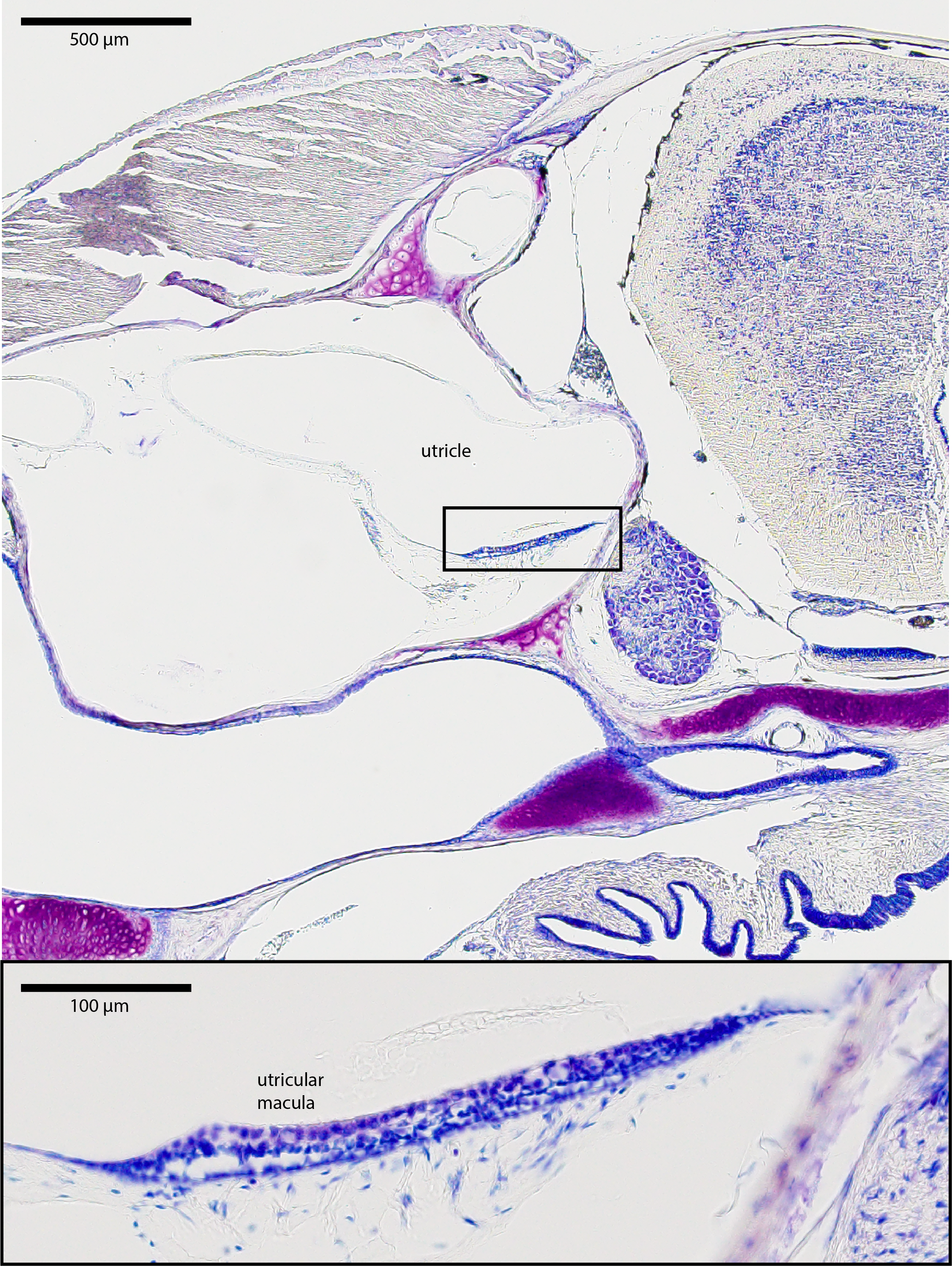
Figure 1. Corresponds to plane b in the scheme, showing the utricle. The insert shows the magnification of the macula, the sensory epithelium of the utricle. The overlying crystals are otoconia, components of the otolithic membrane.
Higher resolution figure 1.
Higher resolution figure 1.
Figure 2. Corresponds to plane c in the scheme, showing the saccule. The insert shows the magnification of the macula of the saccule.
Higher resolution figure 2.
Higher resolution figure 2.
Figure 3. Corresponds to plane d in the scheme, showing the lagenar recess. The insert shows the magnification of the macula of the lagena.
Higher resolution figure 3.
Higher resolution figure 3.


Figure 4. Corresponds to plane e in the scheme, showing the amphibian papilla recess. The insert shows the magnification of the sensory epithelium and the overlying tectorial membrane.
Higher resolution figure 4.
Higher resolution figure 4.
Figure 5. Corresponds to plane f in the scheme, showing the basilar papilla recess. The insert shows the magnification of the sensory epithelium and the overlying tectorial membrane.
Higher resolution figure 5.
Higher resolution figure 5.
Adult frog: NF stage 66.
Figure 1. Corresponds to plane a in the scheme, showing the ampullae of the anterior and horizontal canal. The insert shows the magnification of the anterior ampulla with its crista ampullaris.
Higher resolution figure 1.
Higher resolution figure 1.
Figure 2. Corresponds to plane b in the scheme, showing the utricle. The insert shows the magnification of the macula.
Higher resolution figure 2.
Higher resolution figure 2.
Figure 3. Corresponds to plane c in the scheme, showing the saccule. The insert shows the magnification of the macula of the saccule.
Higher resolution figure 3.
Higher resolution figure 3.

Figure 4. Corresponds to plane d in the scheme, showing the lagenar recess. The insert shows the magnification of the macula of the lagena.
Higher resolution figure 4.
Higher resolution figure 4.
Figure 5. Corresponds to plane e in the scheme, showing the amphibian papilla recess. The insert shows the magnification of the sensory epithelium and the overlying tectorial membrane.
Higher resolution figure 5.
Higher resolution figure 5.
Figure 6. Corresponds to plane f in the scheme, showing the basilar papilla recess. The insert shows the magnification of the sensory epithelium.
Higher resolution figure 6.
Higher resolution figure 6.
Last Updated: 2019-09-01