Click here to close Hello! We notice that you are using Internet Explorer, which is not supported by Xenbase and may cause the site to display incorrectly. We suggest using a current version of Chrome, FireFox, or Safari.
XB-IMG-147689

Xenbase Image ID: 147689

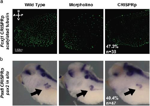
Fig. 3. CRISPR injected embryos phenocopy established mutant or MO knockdown phenotypes in the F0 generation. Left panel=WT embryo, Middle panel=MO injected embryo, Right panel=CRISPRp injected embryo. (a) Foxj1 CRISPRp – Immunohistochemistry of stage 28 embryos with acetylated tubulin to mark surface cilia. (b) Pax8 CRISPRp – in situ hybridization of stage 38 embryos with pax2 probe to stain kidney tubule. Black arrow points to pronephros.

Image published in: Bhattacharya D et al. (2015)

Copyright © 2015. Image reproduced with permission of the Publisher, Elsevier B. V.

GeneSynonymsSpeciesStage(s)Tissue
pax2LOC108697493, pax-2, pax2-a, pax2-b, XPax-2, XPax2X. tropicalisThroughout NF stage 37 and 38otic vesicle
pronephric kidney
brain
midbrain-hindbrain boundary
hindbrain
optic stalk
pronephric nephrostome

Image source: Published

Experiment + Assay Source Phenotypes and Disease
Xtr Wt + foxj1 CRISPR + NF28 (immunohistochemistry) fig.3.a
Disease
facial neoplasm
autoimmune disease
allergic rhinitis
Xtr Wt + foxj1 MO + NF28 (immunohistochemistry) fig.3.a
Disease
facial neoplasm
autoimmune disease
allergic rhinitis
Xtr Wt + pax8 CRISPR + NF37/38 (in situ hybridization) fig.3.b
Anatomical Phenotype
abnormal development of pronephric kidney
abnormal pronephric kidney morphology
Xtr Wt + pax8 MO + NF37/38 (in situ hybridization) fig.3.b
Anatomical Phenotype
abnormal development of pronephric kidney
abnormal pronephric kidney morphology

Larger Image
Printer Friendly View

Return to previous page