XB-IMG-82350
Xenbase Image ID: 82350
|
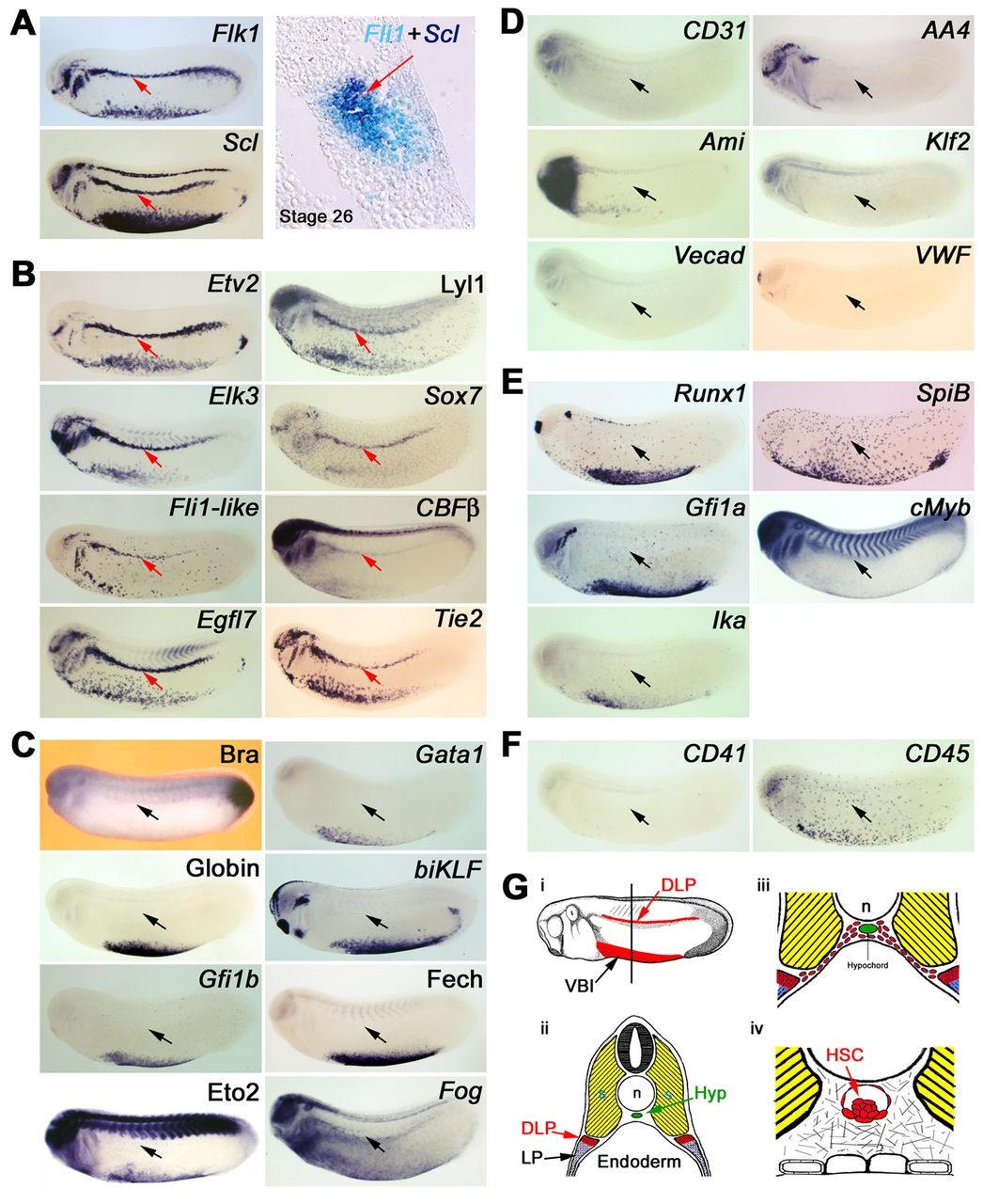
Fig. 1. Characterisation of DLP mesoderm adult haemangioblasts. (A) Adult haemangioblasts localise in the DLP adjacent to the somites and are the earliest HSC progenitors detectable by co-expression of VEGFR2, Flk1, and the stem cell leukaemia gene (Scl), which marks the emergence of haemangioblasts. The panel on the right shows co-expression of the endothelial gene, Fli1, and Scl on a 10 μM section. Note that no morphological differences are observed between adult haemangioblasts and surrounding tissues. (B) Expression analysis revealing novel haematopoietic, Lyl1, Sox7 and CBFβ, as well as novel endothelial, Etv2, Elk3, Fli1-like, Egfl7 and Tie2, gene expression in adult haemangioblasts. (C) Adult haemangioblasts do not express the early mesodermal marker, Brachyury (Bra), nor the erythrocyte differentiation genes, Gata1, Globin, biKLF, Gfi1b, Ferrochelatase (Fech), Eto2 and Fog. (D) Adult haemangioblasts do not express genes associated with mature blood vessels, CD31, AA4, Ami, Klf2, Vecad and VWF. (E) Adult haemangioblasts do not express key HSC-associated genes such as Runx1, SpiB, Gfi1a, cMyb and Ikaros (Ika). (F) Expression of CD41, a gene associated with haematopoietic commitment, and CD45, which is associated with haematopoietic differentiation, are undetectable in adult haemangioblasts. The DLP is indicated by the red or black arrows. All embryos were hybridised as whole mounts and are shown in lateral view, with anterior to the left and dorsal to the top. All embryos are shown at stage 26. (G) Schematic representation of the location of the DLP adult haemangioblast population and its derivatives. (i) Representation of a stage 26 embryo showing in red the Scl expression domains, the ventral blood island and the DLP mesoderm. (ii) Cross section of a stage 26 embryo, at the level indicated by the line in panel i, showing the location of the DLP. Note that adult haemangioblasts (red tissue) lies immediately ventral to the somites (yellow tissue) but at some distance from the hypochord (green tissue), where the DA and HSCs eventually emerge. (iii) Picture showing DA/HSC progenitors migrating from the DLP to the hypochord, a process taking place from stage 28 to 31. (iv) Schematic representation of HSCs emerging in association with the ventral wall of the DA; these haematopoietic clusters are found from stage 42 to 44, 4 days after the specification of adult haemangioblasts in the DLP. Hyp, hypochord; n, notochord; s, somites; VBI, ventral blood island. Image published in: Ciau-Uitz A et al. (2013) Copyright © 2013. Image reproduced with permission of the publisher and the copyright holder. This is an Open Access article distributed under the terms of the Creative Commons Attribution License.
Image source: Published Larger Image Printer Friendly View |