XB-IMG-133068
Xenbase Image ID: 133068
|
|
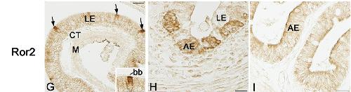
Expression profile of ror2 proteins in the small intestine during natural metamorphosis. Cross sections were immunostained with anti-ror2 (G-H) antibody Ror2 Ab1. Cells positive for Ror2 (G; arrows) are scattered only in the larval epithelium (LE). At NF stage 57, ror2 positive cells are located in the brush border (bb) on the apical surface (see Inset in G). a strong immunoreactivity for Ror2 is localized in islets of the adult epithelium (AE) (H, NF stage 61) LE: larval (foregut) epithelium; AE adult (foregut) epithelium, CT: connective tissue; M: muscles. Scale bars: 20 um. This image is extracted from figure published in: Ishizuya-Oka A et al. (2014), Image published in: Ishizuya-Oka A et al. (2014) Image reproduced on Xenbase with permission of the publisher and the copyright holder. Creative Commons Attribution license Larger Image Printer Friendly View |