XB-IMG-138250
Xenbase Image ID: 138250
|
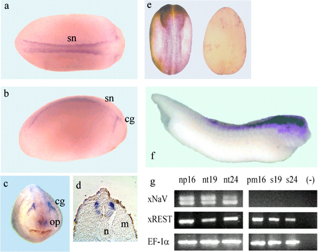
Fig. 1.
NaV1.2 and REST/NRSF are expressed in neural tissues during development of X. laevis.a–d, ISH for xNaV1.2: expression is restricted to primary spinal neurons (sn), cranial ganglia primordia (cg), and olfactory placodes (op). In a section of a stage 18 embryo, xNaV 1.2 expression is observed in the lateral and ventral regions of the neural tube. a, Dorsal view, stage 18; b, lateral view, stage 18;c, anterior view, stage 24; d, central transversal section, stage 18; n, notochord;m, presomitic mesoderm; e,left, ISH showing diffuse expression of REST during neurulation (stage 18), including neural folds; right, a sense REST probe does not produce significant labeling in a stage 18 embryo; f, at stage 35, REST/NRSF expression is stronger in the anterior neural tissue; g, RT-PCR showing the coexpression of xNaV1.2 and REST/NRSF in dissected tissues at the stages annotated; np, neural plate; nt, neural tube; pm, presomitic mesoderm; s, somites. The constitutively expressed transcript EF1α is shown as a control. Image published in: Armisén R et al. (2002) Copyright © 2002. This image is reproduced with permission of the publisher and the copyright holder. This is an Open Access article distributed under the terms of the Creative Commons Attribution License.
Image source: Published Larger Image Printer Friendly View |