XB-IMG-145456
Xenbase Image ID: 145456
|
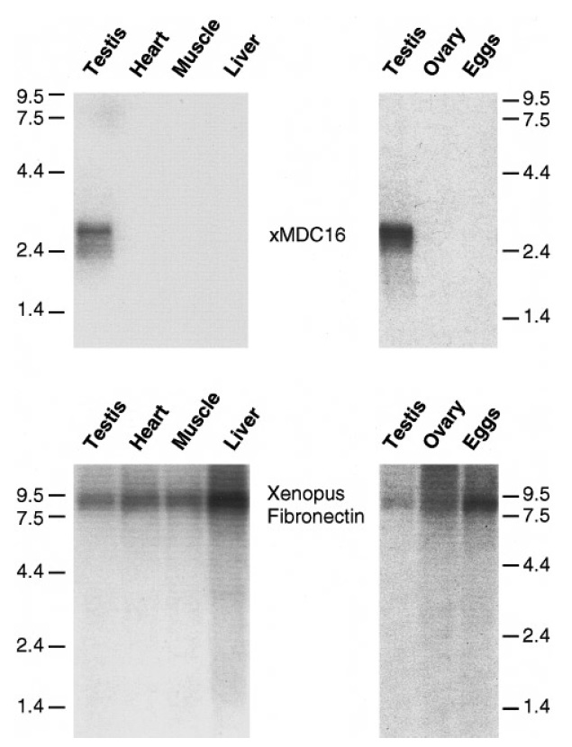
FIG. 3. Northern blot analysis ofxMDC16. The top shows Nordl ern
blots of RNA Isolated from the X. laevls tissues testis, heart,
Uver, and muscle (left) and testis. ovaries, and eggs (right). Both
blots were probed separately w1d1 32P-labeled xMDC16 eDNA under
high stringency conditions (see Methods}. The lower left shows
the same blot as the upper left probed as a control with 32P-labeled
X. Jaev/s fibronectln eDNA (kindly provided by Drs. D. Alfandar1
and D. DeSimone), after removal of the xMDC16 probe. The lower
right shows a Northern blot of RNA samples that were Identical
to dlose shown in the upper right (testis, ovary, and eggs}, probed
with the control 32P-labeled X. Jaev/s fibronectin eDNA. Image published in: Shilling FM et al. (1997) Copyright © 1997. Image reproduced with permission of the Publisher, Elsevier B. V.
Image source: Published Larger Image Printer Friendly View |