XB-IMG-149229
Xenbase Image ID: 149229

|
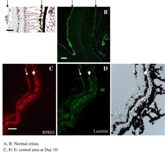
Fig. 3. Laminin and RPE65 distribution in regenerating retinas at Day 10 after retinectomy. (A) Normal Xenopus retina stained with hematoxylin and eosin. Two arrows indicate RVM (retinal vascular membrane) and Bruch's membrane. Both membrane structures are clearly immunoreactive for laminin as indicated by arrows in (B). (C, D, E) Retinectomized eye at Day 10. RPE65, laminin and Nomarsky differential image of the same area at the central (posterior) part of the eye. Arrows and arrowheads indicate the newly formed pigmented epithelium and the original RPE layer, respectively. Both layers are positively stained for RPE65 and laminin. Scale bars in A and B are 30 μm, and bar in C is 20 μm. Image published in: Yoshii C et al. (2007) Copyright © 2007. Image reproduced with permission of the Publisher, Elsevier B. V.
Image source: Published Larger Image Printer Friendly View |
