XB-IMG-158415
Xenbase Image ID: 158415
|
|
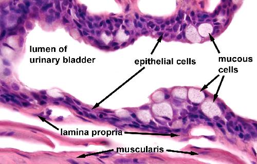
High magnification image of urinary bladder epithelium. The epithelium lining the lumen of the urinary bladder is transitional epithelium. Note the presence of two distinct cell types of the epithelium. The epithelial cells comprise most of the mucous lining and are stratified. Some epithelial cells appear cuboidal in shape, while other appear to be squamous. Interspersed among the epithelial cells are mucus-containing cells that have the same appearance as the flask cells of the collecting tubules of the kidney.
Image from AF Wiechmann and CR Wirsig (2003) "Color Atlas of Xenopus laevis Histology", (page 67, Chapter 6,Urinary system: Figure 22). Copyright 2003. Kluwer Academic Publishers. Reproduced with kind permission from Springer Science & Business Media B.V. Image published in: Color Atlas of Xenopus laevis Histology Larger Image Printer Friendly View |