XB-IMG-158419
Xenbase Image ID: 158419
|
|
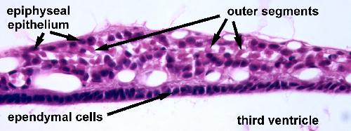
High magnification of the pineal gland. Ependymal cells line the third ventricle. The principal cells of the pineal gland are pinealocytes (also called epiphyseal epithelium), which produce the hormone melatonin. In Xenopus, the pinealocytes have an appearance similar to retinal photoreceptors with outer segments that protrude into the lumens of the gland. Interstitial cells are glial-like cells and are identified by their elongate or irregular-shaped nuclei.
Image from AF Wiechmann and CR Wirsig (2003) "Color Atlas of Xenopus laevis Histology", (page 73, Chapter 7, Endocrine Organs: Figure 10). Copyright 2003. Kluwer Academic Publishers. Reproduced with kind permission from Springer Science & Business Media B.V. Image published in: Color Atlas of Xenopus laevis Histology Larger Image Printer Friendly View |