XB-IMG-43883
Xenbase Image ID: 43883
|
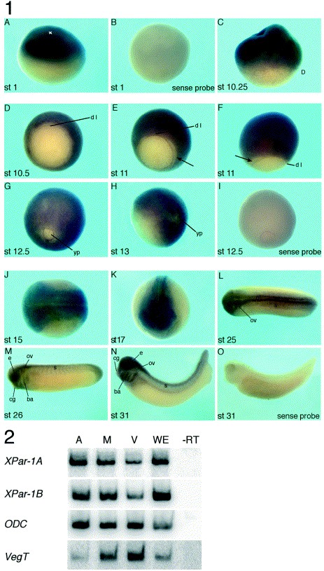
Fig. 2. Whole mount in situ hybridization analysis of XPar-1A expression during embryonic development. (1) Stages of embryogenesis (Nieuwkoop and Faber, 1967) are shown on the left of each panel. (A–C,F,H) Lateral views; (D,E,G,I) vegetal views with dorsal to the top; (K) anterior view; (J,L) dorsal views with anterior to the left; (M–O) lateral views with anterior to the left. ba, branchial arches; cg, cement gland; D, dorsal; dl, dorsal lip of the blastopore; e, eye; ov, otic vesicle; nt, neural tube; s, somites; yp, yolk plug. The cross in (A) indicates animal pole. The arrow in (E,F) marks the gap between the blastopore lip and XPar-1A expression. (2) RT-PCR analysis of XPar-1A and XPar-1B transcripts in animal pole (A), marginal region (M) and vegetal half explants from stage 7 embryos. ODC was used as a loading control. The vegetally localized VegT served as a control for animal–vegetal dissection. Image published in: Ossipova O et al. (2002) Copyright © 2002. Image reproduced with permission of the Publisher, Elsevier B. V.
Image source: Published Larger Image Printer Friendly View |