XB-IMG-46050
Xenbase Image ID: 46050
|
||||||||||
|
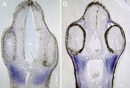
Fig. 6. MMP2 is expressed in tissues that abut the developing visual system. Embryos at stages 32 (A) and 35/36 (B) were processed as wholemounts for in situ hybridization with a digoxigenin-labelled (blue) XMMP2 antisense riboprobe. Vibratome transverse sections (50 μm) through the embryo at the level of the diencephalon. Pigmented cells are shown in black. Broken line represents the ventral diencephalon, with the optic chiasm found at the midline. The arrowheads point to Xmmp2 mRNA-expressing cells that neighbour the optic tectum. R, retina; oc, optic chiasm; Tec, tectum; Ve, ventricle; D, dorsal, V, ventral. Scale bar: 50 μm. Image published in: Hehr CL et al. (2005) Copyright © 2005. Image reproduced with permission of the Publisher and the copyright holder. This is an Open Access article distributed under the terms of the Creative Commons Attribution License.
Image source: Published Larger Image Printer Friendly View |