PCP and Septins Compartmentalize Cortical Actomyosin to Direct Collective Cell Movement
Shindo & Wallingford, published in the journal, Science: 7 February 2014: Vol. 343 no. 6171 pp. 649-652 [DOI: 10.1126/science.1243126]
Despite our understanding of actomyosin function in individual migrating cells, we know little about the mechanisms by which actomyosin drives collective cell movement in vertebrate embryos. The collective movements of convergent extension drive both global reorganization of the early embryo and local remodeling during organogenesis. We report here that planar cell polarity (PCP) proteins control convergent extension by exploiting an evolutionarily ancient function of the septin cytoskeleton. By directing septin-mediated compartmentalization of cortical actomyosin, PCP proteins coordinate the specific shortening of mesenchymal cell-cell contacts, which in turn powers cell interdigitation. These data illuminate the interface between developmental signaling systems and the fundamental machinery of cell behavior and should provide insights into the etiology of human birth defects, such as spina bifida and congenital kidney cysts.
Editor's Choice: Annalisa VanHook, Separate to Intercalate Sci. Signal., 11 February 2014: Vol. 7, Issue 312, p. ec43 [DOI: 10.1126/scisignal.2005164]
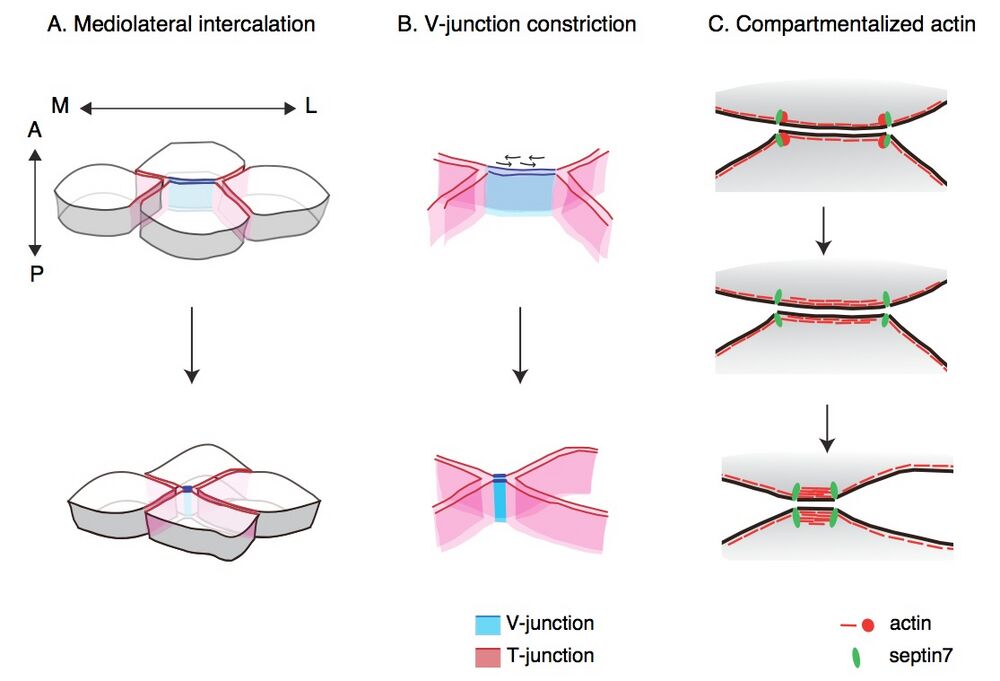
Planar cell polarity (PCP) signaling establishes directionality in epithelial sheets and is important for various morphogenetic events, including convergent extension (CE) in gastrulating vertebrate embryos. During CE, mediolateral cells move toward the dorsal midline and intercalate to extend the body axis, and neural tube defects such as spina bifida result when CE is disrupted. Shindo and Wallingford investigated the link between PCP and cell shape change during intercalation of the mesoderm in gastrulating Xenopus (frog) embryos. As these approximately hexagonal cells intercalated, the anterior and posterior edges of each cell aligned roughly perpendicular to the embryonic midline, and the boundaries between the posterior face of one cell and the anterior face of a neighboring cell shortened and became enriched in phosphorylated myosin II (pMyoII). Actin accumulated at the vertices between the anteroposterior boundaries and the mediolateral sides of the cells (the mediolateral vertices), and cortical actin flowed along the anteroposterior boundaries but not along the mediolateral edges. Whereas mediolateral edges were under constant tension, the anteroposterior boundaries became increasingly taut as they shortened. Septins are GTP-binding scaffold proteins that form filaments involved in cytoskeletal dynamics, including PCP-directed cell migration and ciliogenesis. Fluorescently tagged Septin 7 (Sept7) was enriched at mediolateral vertices, and knocking down Sept7 prevented actin accumulation at these sites, blocked the tensioning of anteroposterior boundaries, increased the abundance of pMyoII, and eliminated the polar distribution of pMyoII. Knocking down the core PCP component Dishevelled interfered with the polar distribution of both Sept7 and pMyoII and decreased the abundance of pMyoII. pMyoII was required for tensioning of the anteroposterior boundaries and for normal CE. These results suggest a model in which the PCP network, which promotes the phosphorylation of MyoII, also promotes the localization of Sept7 to mediolateral vertices, thus creating separate cortical actomyosin compartments, allowing the anteroposterior actomyosin network to contract to drive intercalation.
From Shindo & Wallingford, Science: 7 February 2014: Vol. 343 no. 6171 pp. 649-652 [DOI: 10.1126/science.1243126]. Reprinted with permission from AAAS.
homepage slider image: Immunostaining of pMyo and membrane in notochord, by Asako Shindo
Last Updated: 2014-04-23

