Basal cells in Xenopus mucociliary epithelium
ΔN-Tp63 Mediates Wnt/β-Catenin-Induced Inhibition of Differentiation in Basal Stem Cells of Mucociliary Epithelia
Cell Reports VOLUME 28, ISSUE 13, P3338-3352.E6, SEPTEMBER 24, 2019. https://doi.org/10.1016/j.celrep.2019.08.063.
Maximilian Haas, Jose Luis Gomez Vazquez, Dingyuan Iris Sun, Hong Thi Tran, Magdalena Brislinger, Alexia Tasca, Orr Shomroni, Kris Vleminckx, and Peter Walentek.
Impaired (re-)generation of lung epithelia is associated with Wnt signaling changes in animals and human lung disease patients. Haas et al. demonstrate that ΔN-Tp63 is a Wnt-regulated master transcription factor inhibiting (re-) generation of new epithelial cells from stem cells. These findings are equally important for understanding animal development and disease mechanisms.
Highlights
- ΔN-Tp63 is a master regulator of stemness versus differentiation in mucociliary cells
- Wnt/β-catenin inhibits differentiation by upregulating ΔN-Tp63 in basal stem cells
- Wnt/ΔN-Tp63-induced mucociliary remodeling is reversible in vivo and in vitro
- The Xenopus epidermis provides a model to study basal stem cells in vivo
Click here to view article at Cell Reports.
Click here to view article at Pubmed.
Click here to view article on Xenbase.
SUMMARY
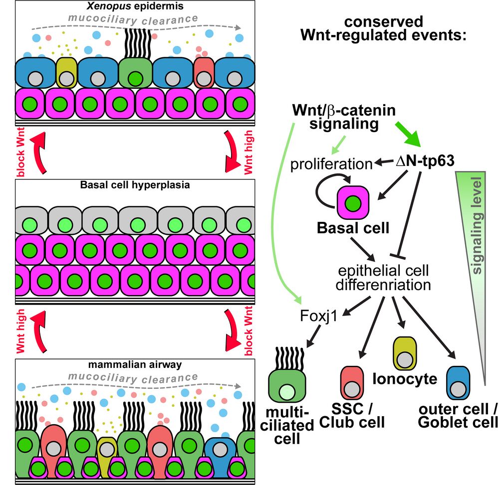
Mucociliary epithelia provide a first line of defense against pathogens. Impaired regeneration and re- modeling of mucociliary epithelia are associated with dysregulated Wnt/b-catenin signaling in chronic airway diseases, but underlying mechanisms remain elusive, and studies yield seemingly contradicting results. Employing the Xenopus mucociliary epidermis, the mouse airway, and human airway basal cells, we characterize the evolutionarily conserved roles of Wnt/beta-catenin signaling in vertebrates. In multiciliated cells, Wnt is required for cilia formation during differentiation. In basal cells, Wnt prevents specification of epithelial cell types by activating deltaN-TP63, a master transcription factor, which is necessary and sufficient to mediate the Wnt-induced inhibition of specification and is required to retain basal cells during development. Chronic Wnt activation leads to remodeling and basal cell hyperplasia, which are reversible in vivo and in vitro, suggesting Wnt inhibition as a treatment option in chronic lung diseases. Our work provides important insights into mucociliary signaling, development, and disease.
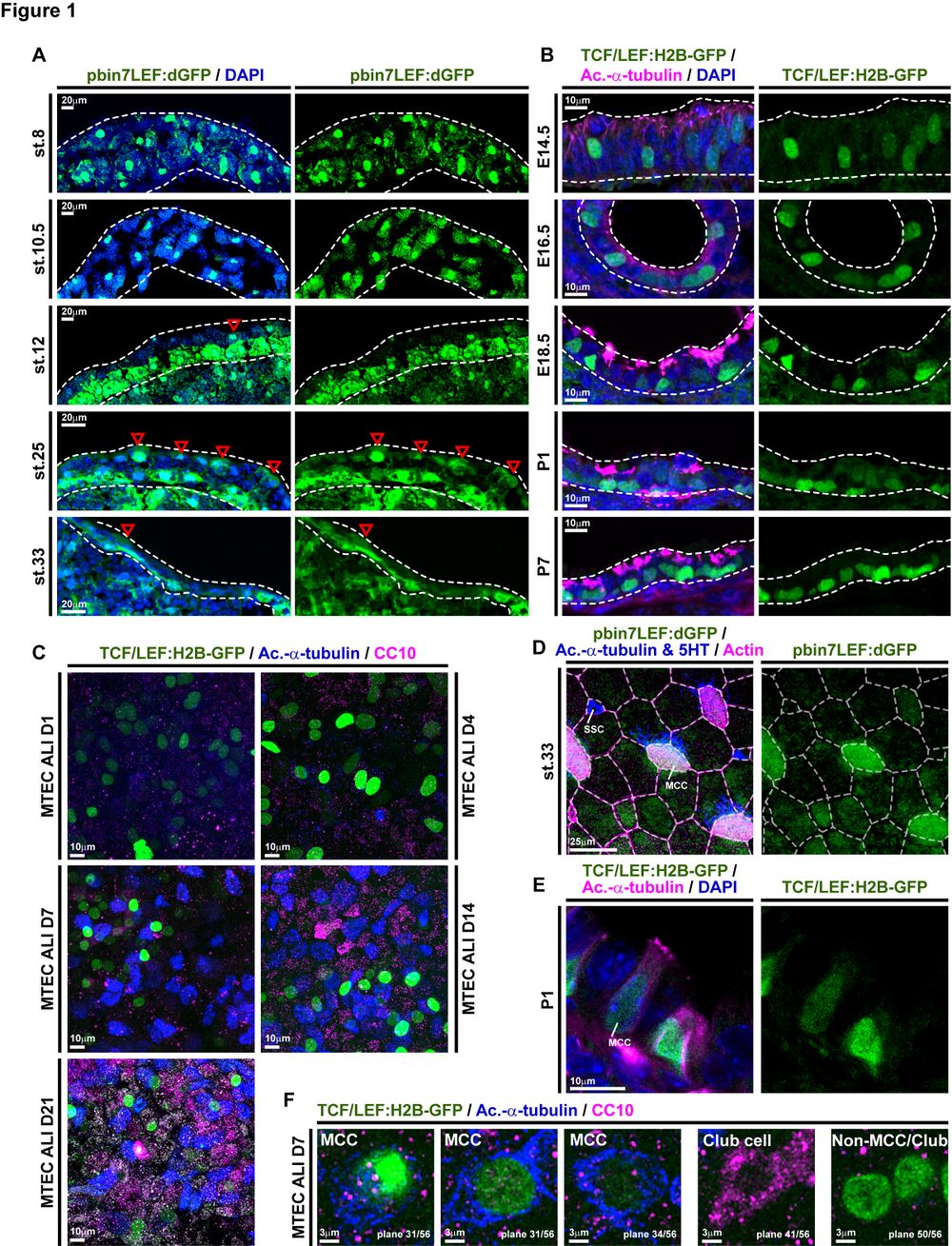
Figure 1. Wnt/b-Catenin Signaling Is Active in MCCs and Basal Progenitors
(A) Analysis of Wnt/b-catenin activity in the X. laevis mucociliary epidermis using the pbin7LEF:dGFP reporter line (green). Nuclei are stained by DAPI (blue). Red arrowheads indicate GFP-positive cells in the outer epithelial layer. Dashed lines outline the epidermal layers. Embryonic stages (st. 8–33) are indicated.
(B) Analysis of Wnt/b-catenin activity in the mouse developing airway mucociliary epithelium using the TCF/LEF:H2B-GFP reporter line (green). Nuclei are stained by DAPI (blue) and MCCs are marked by acetylated-a-tubulin (Ac.-a-tubulin, magenta) staining. Dashed lines outline the epithelium. Embryonic (E14.5–18.5) and post-natal (P1–7) stages are indicated.
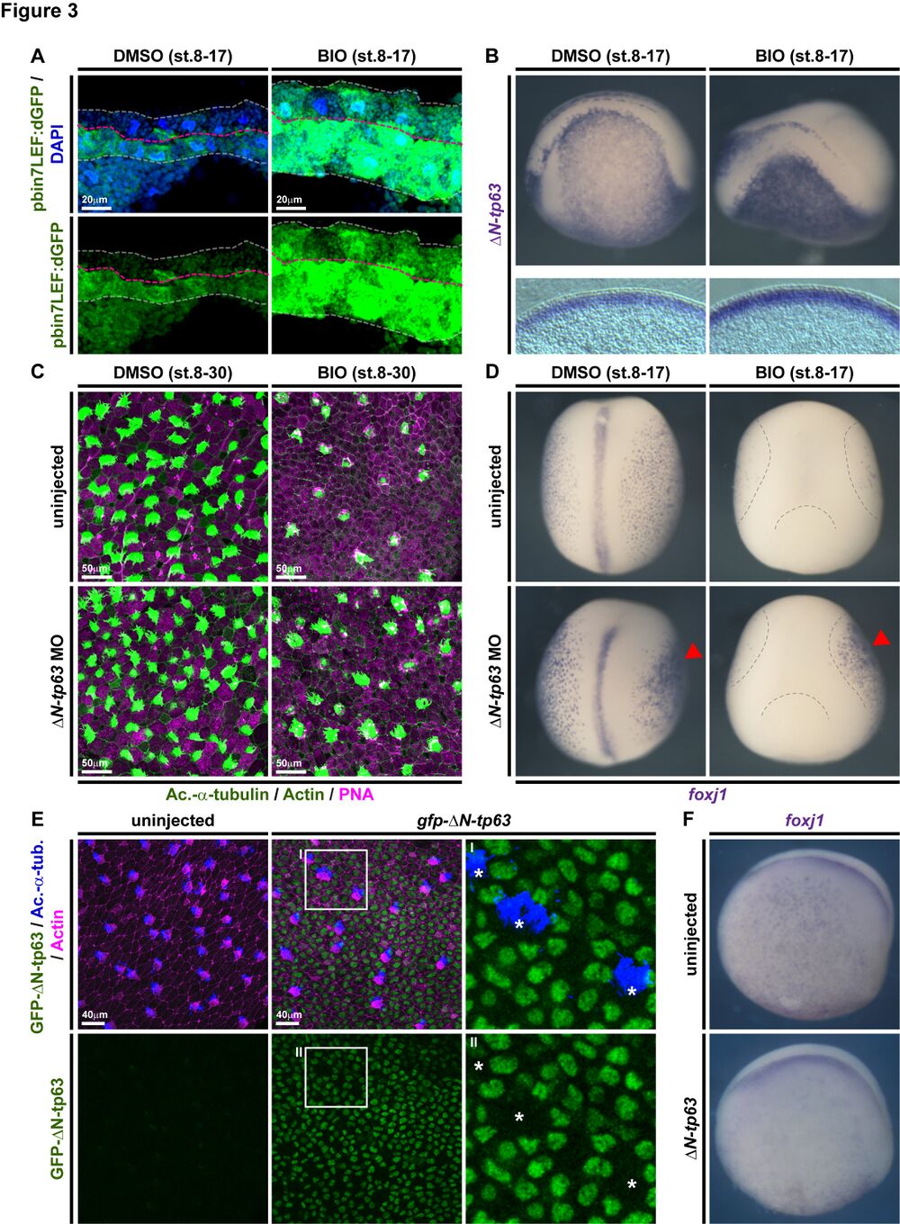
Figure 3. DN-tp63 Mediates Wnt/b-Catenin-Induced Inhibition of Cell Fate Specifi- cation in Xenopus
(A–D) BIO treatments from st. 8–17 or st. 8–30. DMSO was used as vehicle control.
(A) Confocal imaging shows Wnt/b-catenin signaling activation (green) and thickening of the epidermis in BIO-treated embryos. Nuclei (DAPI, blue). DMSO (n = 2); BIO (n = 2).
(B) In situ hybridization (ISH) shows increased in- tensity and thickness of the DN-tp63 expression domain in BIO-treated whole mounts (upper row) and transversal sections (bottom row).
(C) BIO treatment reduces MCC (Ac.-a-tubulin, green), ionocyte (no staining, black), and SSC (large vesicles, peanut agglutinin [PNA] staining, magenta) numbers in confocal micrographs. Actin staining (green). DN-tp63 MO in controls leads to increased MCCs and ionocytes, while DN-tp63 MO in BIO-treated embryos rescues MCC and ionocyte formation.
(D) ISH reveals reduced MCC numbers (foxj+ cells) after BIO treatment, while unilateral knockdown of DN-tp63 in control treated embryos leads to more foxj+ cells and rescues foxj+ cell numbers in BIO- treated embryos. DMSO (n = 5); BIO (n = 7); DMSO+DN-tp63 MO (n = 5); BIO+DN-tp63 MO (n = 4). Injected side is indicated by red arrowhead. Dashed lines indicate epidermal area in BIO- treated embryos.
(E) Overexpression of gfp-DN-tp63 mRNA leads to nuclear localization of the protein (green) and reduced MCC (Ac.-a-tubulin, blue) numbers at st. 30. Actin staining (magenta). Differentiated MCCs in injected specimens show no nuclear GFP signal (asterisks), indicating that they were not targeted. Magnified areas I–II are indicated.
(F) ISH for foxj1 at st. 17 shows reduced MCCs in DN-tp63 mRNA injected embryos.
Related to Figures S2 and S3.
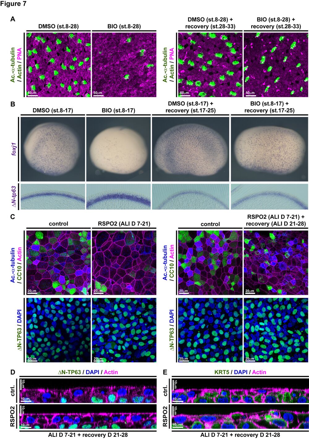
Figure 7. Wnt/b-Catenin-Induced Increase in BCs and Loss of Epithelial Differentiation Are Reversible
(A and B) In Xenopus, BIO treatments from st. 8–17 or st. 8–30 inhibit differentiation as compared to DMSO treated controls, but the epithelium can regenerate after removal of BIO and recovery until st. 33 (A) or st. 25 (B).
(A) BIO treatment reduces MCC (Ac.-a-tubulin, green), ionocyte (no staining, black), and SSC (large vesicles, PNA staining, magenta) numbers in confocal micrographs at st. 28, which recover af- ter regeneration until st. 33. Actin staining (green). (B) ISH shows reduction in foxj1 expressing cells and an increase in DN-tp63 expression in BIO- treated whole mounts (upper row) and transversal sections (bottom row) at st. 17, which both recover after regeneration until st. 25. DMSO (n = 5); BIO st. 17 (n = 5); DMSO st. 25 (n = 5); BIO+recovery st. 25 (n = 5).
(C) Confocal imaging of specimens stained for Ac.-a-tubulin (MCCs, blue), CC10 (club cells, green), and actin (cell membranes, magenta) show reduced MCC and club cell numbers in RSPO2- treated cultures from ALI D7 to D21 but regener- ation of MCCs and club cells at ALI D28. n = 3 cultures per time point and treatment (upper panels). No loss of DN-TP63+ (green) BCs is observed in these experiments (bottom panels). Nuclei (DAPI, blue).
(D and E) Optical orthogonal sections of confocal images. RSPO2-treated cultures display normal- ized epithelial thickness and staining for BC markers DN-TP63 (green, in D) and KRT5 (green, in E) after regeneration at ALI D28.
Related to Figures S6 and S7.
Adapted with permission from Cell Press on behalf of Cell Reports: Haas et al. (2019). ΔN-Tp63 Mediates Wnt/β-Catenin-Induced Inhibition of Differentiation in Basal Stem Cells of Mucociliary Epithelia. Cell Reports VOLUME 28, ISSUE 13, P3338-3352.E6, SEPTEMBER 24, 2019. https://doi.org/10.1016/j.celrep.2019.08.063.
This work is licensed under a Creative Commons Attribution 4.0 International License. The images or other third party material in this article are included in the article’s Creative Commons license, unless indicated otherwise in the credit line; if the material is not included under the Creative Commons license, users will need to obtain permission from the license holder to reproduce the material. To view a copy of this license, visit http://creativecommons.org/licenses/by/4.0/
Last Updated: 2019-09-24