Predation threats over 24 hours activate extension of axons in tadpole brains
Predation threats for a 24-h period activated the extension of axons in the brains of Xenopus tadpoles
Mori T , Kitani Y , Hatakeyama D , Machida K , Goto-Inoue N , Hayakawa S , Yamamoto N , Kashiwagi K , Kashiwagi A.
Sci Rep July 16, 2020; 10 (1): 11737.
Click here to view article at Scientific Reports.
Click here to view article on Pubmed.
Click here to view article on Xenbase.
Abstract
The threat of predation is a driving force in the evolution of animals. We have previously reported that Xenopus laevis enhanced their tail muscles and increased their swimming speeds in the presence of Japanese larval salamander predators. Herein, we investigated the induced gene expression changes in the brains of tadpoles under the threat of predation using 3''-tag digital gene expression profiling. We found that many muscle genes were expressed after 24 h of exposure to predation. Ingenuity pathway analysis further showed that after 24 h of a predation threat, various signal transduction genes were stimulated, such as those affecting the actin cytoskeleton and CREB pathways, and that these might increase microtubule dynamics, axonogenesis, cognition, and memory. To verify the increase in microtubule dynamics, DiI was inserted through the tadpole nostrils. Extension of the axons was clearly observed from the nostril to the diencephalon and was significantly increased (P ≤ 0.0001) after 24 h of exposure to predation, compared with that of the control. The dynamic changes in the signal transductions appeared to bring about new connections in the neural networks, as suggested by the microtubule dynamics. These connections may result in improved memory and cognition abilities, and subsequently increase survivability.
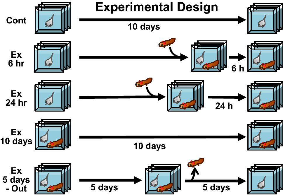
Figure 1.
Experimental design for inducing predation in Xenopus tadpoles. Xenopus tadpoles were assigned to 5 different treatment groups. The control group (Cont) was not exposed to the salamander predator during the experiment (10 days). Tadpoles in the 6 h and 24 h groups were exposed to a salamander larva for 6 or 24 h before sampling, respectively. The 10-day group was exposed to a salamander larva for 10 full days, while the 5-days out group was exposed to a salamander larva for 5 days, to induce the predation threat, then the salamander was removed, and the tadpoles were kept for a further 5 days without the predation threat.
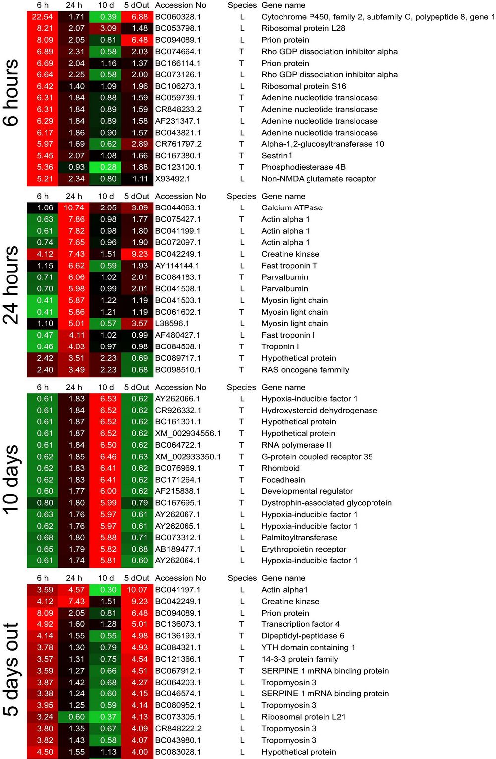
Figure 2.
Heat map using 3′-tag digital gene expression profiles from Xenopus laevis whole brain tissues. Gene expression profiling was performed using the top 15 genes that were expressed by over 1,000 tags from each of the experimental groups described in Fig. 1. The numbers on the heat map indicate the total number of tags expressed for each gene in the experimental and control groups. Accession No: gene number obtained from the NCBI database. Species: “L” indicates Xenopus laevis, and “T” indicates Xenopus tropicalis. The heat map colors and numbers indicate the fold changes in gene expression compared with that of the control.
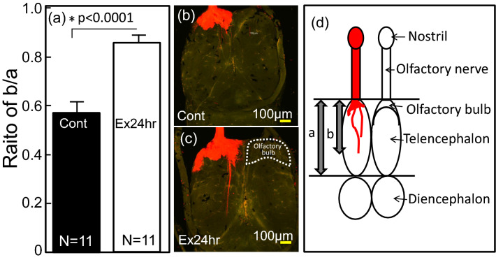
Figure 8.
Extension of the axons in the brains of Xenopus laevis tadpoles exposed to the Japanese predatory larval salamanders, Hynobius lichenatus for 24 h. (a) Ratio of the extended axons per telencephalon compared with the control (Cont.; not exposed to Japanese predators) was calculated as b/a (a: length of telencephalon; b: length of the extended axons) in the brain. Statistical analysis (t-test) on the data were performed with SPSS. (b) Tadpoles not exposed to a predator (c) Tadpoles exposed to Japanese predator for 24 h. (d) Schematic profile of a tadpole’s brain stained by DiI.
Adapted with permission from Nature Publishing Group on behalf of Scientific Reports: Mori et al. (2020). Predation threats for a 24-h period activated the extension of axons in the brains of Xenopus tadpoles. Sci Rep. 2020 Jul 16;10(1):11737. doi: 10.1038/s41598-020-67975-7.
This work is licensed under a Creative Commons Attribution 4.0 International License. The images or other third party material in this article are included in the article’s Creative Commons license, unless indicated otherwise in the credit line; if the material is not included under the Creative Commons license, users will need to obtain permission from the license holder to reproduce the material. To view a copy of this license, visit http://creativecommons.org/licenses/by/4.0/
Last Updated: 2020-08-13