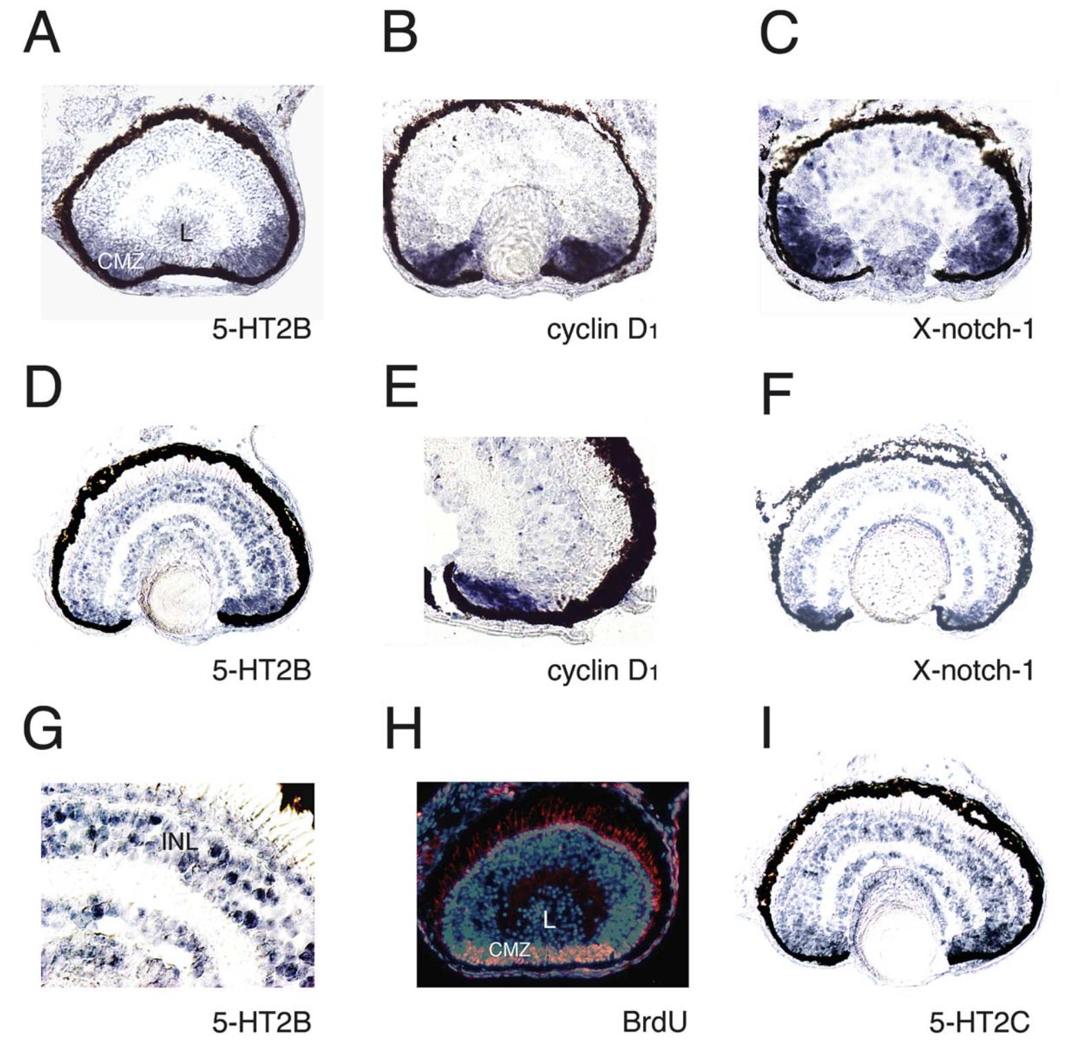
Fig. 4. Expression of 5-HT2B and 2C receptor mRNAs in developing retina. Transverse sections through the eye of a stage 40 (AâC) and 45 larva (DâI). The probes used are indicated at bottom of each panel. The CMZ is labelled by proliferation markers (B,C,E,F) and stained with anti-BrdU antibody after BrdU incorporation (H). In blue, Hoechst nuclear staining; orange, BrdU incorporation. (G) is an enlargement of (D) to show the scattered postmitotic cells expressing 5-HT2B transcripts in the INL. CMZ, ciliary marginal zone; L, lens; INL, inner nuclear layer.
Image published in: De Lucchini S et al. (2003)
Copyright © 2003. Image reproduced with permission of the Publisher.
| Gene | Synonyms | Species | Stage(s) | Tissue |
|---|---|---|---|---|
| htr2b.L | 5-HT2B, LOC108718039 | X. laevis | Throughout NF stage 40 | retina lens eye ciliary marginal zone |
| ccnd1.L | ccnd1-a, ccnd1-b, cyclind1, parathyroid adenomatosis 1, prad1 | X. laevis | Throughout NF stage 40 | retina eye ciliary marginal zone |
| notch1.L | notch, notch-1, xnotch, x-notch-1, xnotch1, xotch | X. laevis | Throughout NF stage 40 | eye retina ciliary marginal zone |
| htr2b.L | 5-HT2B, LOC108718039 | X. laevis | Throughout NF stage 45 | retina ciliary marginal zone eye retinal inner nuclear layer |
| ccnd1.L | ccnd1-a, ccnd1-b, cyclind1, parathyroid adenomatosis 1, prad1 | X. laevis | Throughout NF stage 45 | retina ciliary marginal zone eye |
| notch1.L | notch, notch-1, xnotch, x-notch-1, xnotch1, xotch | X. laevis | Throughout NF stage 45 | eye retina ciliary marginal zone |
| htr2c.S | 5-HT2C | X. laevis | Throughout NF stage 45 | retina ciliary marginal zone eye retinal inner nuclear layer |
Image source: Published
Permanent Image Page
Printer Friendly View
XB-IMG-171071