| Images | Sources | Experiment + Assay | Phenotypes | Human Diseases | ||||
|---|---|---|---|---|---|---|---|---|
|
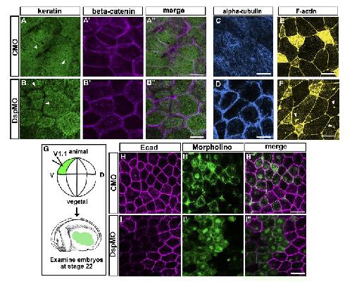
Fig. 6 D
Bharathan NK and Dickinson AJG (2019) |
Xla Wt + dsp MO
NF19-20 (immunohistochemistry) |
|
skin disease
|
||||
|
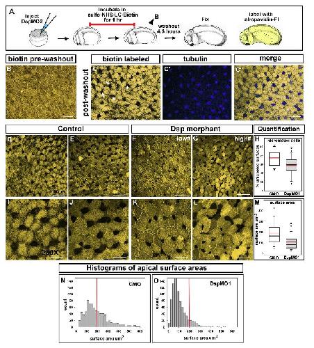
Fig. 5 F G H K L M
Bharathan NK and Dickinson AJG (2019) |
Xla Wt + dsp MO
NF19-20 (immunohistochemistry) |
|
skin disease
|
||||
|
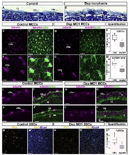
Fig. 4 B
Bharathan NK and Dickinson AJG (2019) |
Xla Wt + dsp MO
NF26-28 (histology) |
|
skin disease
|
||||

|
Fig. 4 D E
Bharathan NK and Dickinson AJG (2019) |
Xla Wt + dsp MO
NF26-28 (immunohistochemistry) |
|
skin disease
|
||||

|
Fig. 4 G H
Bharathan NK and Dickinson AJG (2019) |
Xla Wt + dsp MO
NF26-28 (immunohistochemistry) |
|
skin disease
|
||||

|
Fig. 4 K K'
Bharathan NK and Dickinson AJG (2019) |
Xla Wt + dsp MO
NF26-28 (immunohistochemistry) |
|
skin disease
|
||||

|
Fig. 4 L L'
Bharathan NK and Dickinson AJG (2019) |
Xla Wt + dsp MO
NF26-28 (immunohistochemistry) |
|
skin disease
|
||||

|
Fig. 4 N N' O
Bharathan NK and Dickinson AJG (2019) |
Xla Wt + dsp MO
NF26-28 (immunohistochemistry) |
|
skin disease
|
||||
|
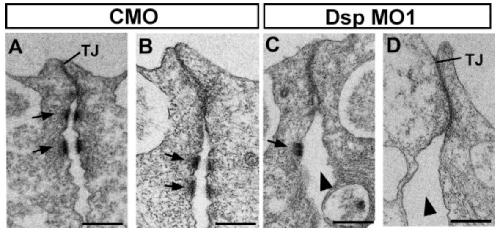
Fig. S 3 C D
Bharathan NK and Dickinson AJG (2019) |
Xla Wt + dsp MO
NF26-28 (transmission electron microscopy) |
|
skin disease
|
||||
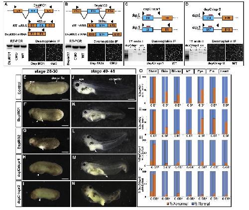
|
Fig. 2 F
Bharathan NK and Dickinson AJG (2019) |
Xla Wt + dsp MO
NF28-29/30 (whole-mount microscopy) |
|
skin disease
|