XB-IMG-131069
Xenbase Image ID: 131069
|
|
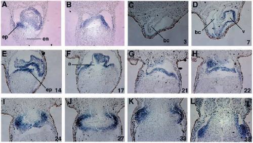
Fig. 3. Serial sections through
the heart of a stage 35 (A,B) and
a stage 40 (C-L) embryo stained
for XTbx5 mRNA expression.
(A) Section through the ventricle
of a stage 35 embryo.
Expression is detected in both
the endocardium and
myocardium as well as in the
tissue we identify as epicardium
(ep), overlying the endoderm
(en). (B) Section through the
sinus venosus slightly posterior
to the section in A. XTbx5
mRNA is detected in the ventral
part of the sinus venosus. (CL)
Selected serial sections
through the heart of a stage 40
embryo proceeding from
anterior to posterior. (C) Bulbus
cordis (bc) (D) Bulbus cordis
(bc) and ventricle (v). XTbx5 is
expressed only in the ventricle.
(E) Ventricle. Expression is also detected in the underlying epicardium (ep). (F,G) Sections through the atrium (a) as it turns dorsally.
(H-L) Sections through the developing sinus venosus. The sections through the heart in C-L have been numbered going from anterior to
posterior, beginning with the bulbus cordis and ending with the sinus venosus. The corresponding number for each section is printed in the
lower right-hand corner of each panel. Each section is 14 mm thick. The bulbus cordis encompasses sections 1-8 (112 mm), the ventricle
sections 5-16 (168 mm), the atrium sections 17-22 (84 mm) and the sinus venosus sections 23-46 (336 mm). Image published in: Horb ME and Thomsen GH (1999) Copyright © 1999. Image reproduced with permission of the Publisher and the copyright holder. This is an Open Access article distributed under the terms of the Creative Commons Attribution License. Larger Image Printer Friendly View |