XB-IMG-131072
Xenbase Image ID: 131072
|
|
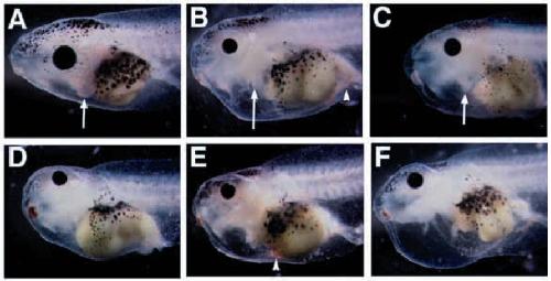
Fig. 6. Embryonic phenotypes generated by a dominant negative XTbx5
protein. (A) Wild-type stage 45 embryo. Arrow points to the heart. Notice how
the heart extends to the edge of the pericardial cavity and is near the ventral
surface of the gut. (B-F) Typical phenotypes caused by XTbx5-EnR-GR. In each
case, embryos were injected dorsally at the 4-cell stage with 1 ng of mRNA and
treated with dexamethasone at stage 14/15. (B,C) Examples of the reduced
heart phenotype. Arrow points to the small heart present in this class of
embryo. (D-F) Examples of the beating nub or heartless phenotype. This
phenotype is typified by the absence of a morphologically recognized heart.
Sometimes, some small amount of beating tissue is present in the dorsal portion
of the pericardial cavity. In B-F, notice the bloated pericardial cavity and
improper gut formation in the injected embryos. In addition, the eyes are
smaller in size. Arrowhead in B and E points to regions where blood cells have
pooled. Image published in: Horb ME and Thomsen GH (1999) Copyright © 1999. Image reproduced with permission of the Publisher and the copyright holder. This is an Open Access article distributed under the terms of the Creative Commons Attribution License. Larger Image Printer Friendly View |